- leptomeninges = arachnoid and pia
advertisement
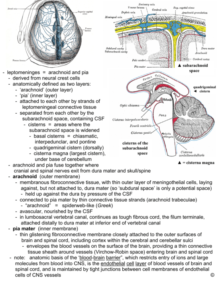
▲ subarachnoid space - leptomeninges = arachnoid and pia - derived from neural crest cells - anatomically defined as two layers: quadrigeminal - ‘arachnoid’ (outer layer) ◄ cistern - ‘pia’ (inner layer) - attached to each other by strands of leptomeningeal connective tissue - separated from each other by the subarachnoid space, containing CSF - cisterns = areas where the subarachnoid space is widened - basal cisterns = chiasmatic, interpedunclar, and pontine cisterns of the - quadrigeminal cistern (dorsally) subarachnoid space - cisterna magna (largest cistern), under base of cerebellum ▲ = cisterna magna - arachnoid and pia fuse together where cranial and spinal nerves exit from dura mater and skull/spine - arachnoid (outer membrane) - membranous fibroconnective tissue, with thin outer layer of meningothelial cells, laying against, but not attached to, dura mater (so ‘subdural space’ is only a potential space) - held up against the dura by pressure of the CSF - connected to pia mater by thin connective tissue strands (arachnoid trabeculae) - “arachnoid” = spiderweb-like (Greek) - avascular, nourished by the CSF - in lumbosacral vertebral canal, continues as tough fibrous cord, the filum terminale, attached distally to dura mater at inferior end of vertebral canal - pia mater (inner membrane) - thin glistening fibroconnective membrane closely attached to the outer surfaces of brain and spinal cord, including cortex within the cerebral and cerebellar sulci - envelopes the blood vessels on the surface of the brain, providing a thin connective tissue sheath around vessels (Virchow-Robin space) entering brain and spinal cord - note: anatomic basis of the ‘blood-brain barrier”, which restricts entry of ions and large molecules from blood into CNS, is the endothelial cell layer of blood vessels of brain and spinal cord, and is maintained by tight junctions between cell membranes of endothelial cells of CNS vessels ©
