Microbiology Mid-Term Exam Review Guide
advertisement
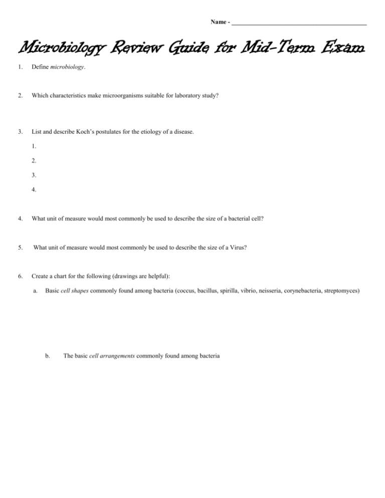
Name - ___________________________________________ Microbiology Review Guide for Mid-Term Exam 1. Define microbiology. 2. Which characteristics make microorganisms suitable for laboratory study? 3. List and describe Koch’s postulates for the etiology of a disease. 1. 2. 3. 4. 4. What unit of measure would most commonly be used to describe the size of a bacterial cell? 5. What unit of measure would most commonly be used to describe the size of a Virus? 6. Create a chart for the following (drawings are helpful): a. Basic cell shapes commonly found among bacteria (coccus, bacillus, spirilla, vibrio, neisseria, corynebacteria, streptomyces) b. The basic cell arrangements commonly found among bacteria 7. Define / describe each of the following as they apply to microbiology: a. Culture b. Inoculum c. Inoculation d. Colony e. Growth medium (-ia) f. Pure culture g. Contaminated culture 8. Describe the procedure for making a bacterial smear for microscopic observation (don’t forget aseptic technique!). 9. Why should a bacterial smear be air-dried before heat fixing (“flaming”) it? What is the purpose/effect of flaming a bacterial smear slide? 10. Describe the procedure for making a gram stain (don’t forget aseptic technique!). 11. Describe the four Biosafety Levels (BSL) 12. What is epidemiology? 13. Differentiate between epidemic and pandemic. 14. Describe how microorganisms cause disease. 15. Describe how antibiotics work. 16. Describe the action of the following antibiotics: a. penicillin b nystatin c rifampin d tetracycline 17. Describe the six (6) ways a pathogen can enter the body. 18. What does the term nosocomial mean? 19. What are zoonoses? 20. Define the phrase “index case” or “patient zero”. 21. Name the outbreak that for which the people listed below have been identified as “patient zero”: Mary Mallon Mabalo Lokela Liu Jianlun A baby in the Lewis House at 40 Broad Street 22. Draw and label the five (5) oxygen relationships. 23. Give the function of each of the following: Leukocidins Hemolysins Coagulases Streptokinase Hyaluronidase Exotoxins Neurotoxins Enterotoxins Endotoxins Exoenzyme 24 Differentiate between the following colony characteristics on an agar plate: a. Form b. Elevation c. Margin d. Surface e. Opacity f. Chromogenesis 25. Label the following colony characteristics: 26. Differentiate mesophile, psychrophile and thermophile 27. Differentiate acidophile and alkalinophile. 28. Write out and explain the reactions catalyzed by the following enzymes: Superoxide dismutase Catalase 29. Explain the importance of the endospore and diagram the six endospore morphologies. 30. Define the following and provide an example: Bacteriology Virology Mycology Parasitology Protozoology Phycology 31. Complete the following table of units of measure commonly used in microbiology. Unit 1 centimeter 1 nanometer Abbreviation Exponential Value cm mm 10-3 μm 10-6 nm 32. Draw and label diagrams comparing the structure and composition of a Gram positive cell wall to that of a Gram negative cell wall. (Use colored pencils) 33. What three things should you do immediately upon entering the lab? 34. What three things should you do before you leave the lab? 35. What is aseptic technique? 36. Explain how to flame sterilize a loop. 37. Explain how you prepare a streak plate. 38. What is the purpose of making a streak plate? 39. Explain how you begin to focus on a prepared slide. 40. Why isn’t the coarse focus used beyond the 10X objective lens? 41. In preparing a slide for simple staining, why is it important to let the drop dry completely? 42. What is the purpose of heat fixing? 43. Why is crystal violet used in the Gram staining procedure? 44. Why is iodine used in the Gram staining procedure? 45. Why is alcohol used after the iodine is used, but not after the crystal violet step? 46. What is the purpose of the negative stain? 47. Describe the steps in a negative stain. 48. Describe the steps in an endospore stain. And explain why the endospore stain requires heating. 49. Draw and label a typical growth curve. 50. Differentiate the following modes of nutrition: Chemoheterotroph Chemoautotroph Photoheterotroph Photoautotroph Make a chart for our “meet the microbes” and “meet the microbe hunters” units. Use the space below to illustrate the proper labeling of an agar plate (using Bacillus subtilis, Nutrient agar, and 37ºC)
