Know About
advertisement
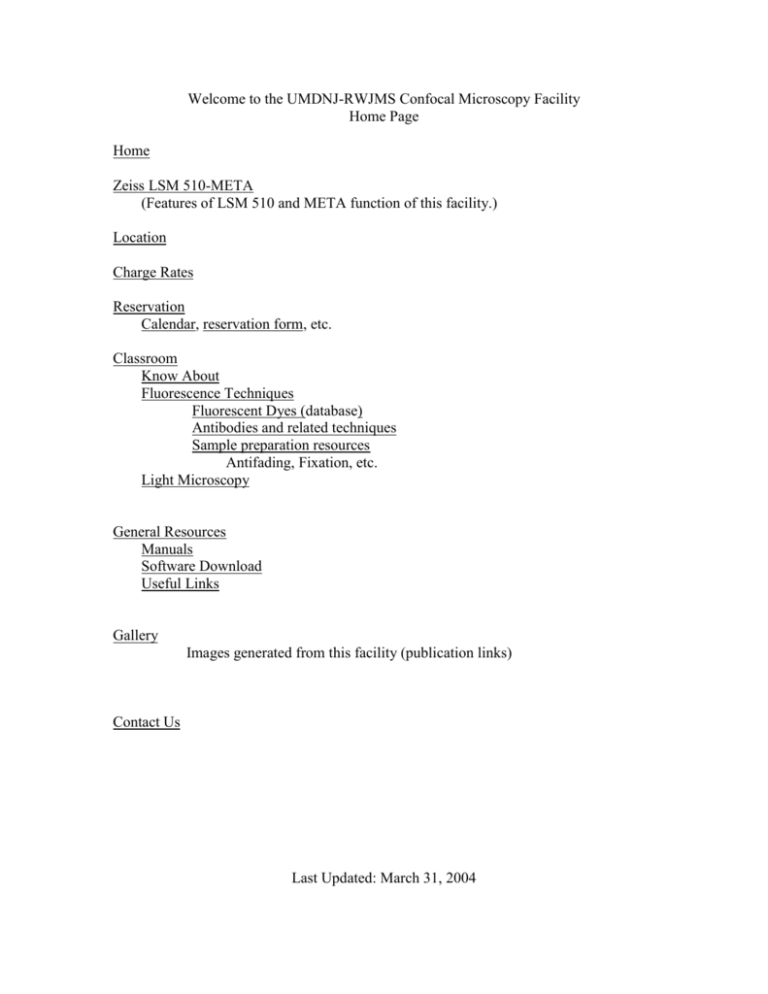
Welcome to the UMDNJ-RWJMS Confocal Microscopy Facility Home Page Home Zeiss LSM 510-META (Features of LSM 510 and META function of this facility.) Location Charge Rates Reservation Calendar, reservation form, etc. Classroom Know About Fluorescence Techniques Fluorescent Dyes (database) Antibodies and related techniques Sample preparation resources Antifading, Fixation, etc. Light Microscopy General Resources Manuals Software Download Useful Links Gallery Images generated from this facility (publication links) Contact Us Last Updated: March 31, 2004 Home This latest state-of-art Zeiss LSM 510-META confocal microscope is equipped with UV and three visible light lasers, three fluorescence detectors including one with polychromatic function and a transmitted light detector. The Axiovert 200 inverted Scope used by the system has 10, 20 (DIC) and 63X (DIC) water immersion objectives, which is perfect for live cell imaging. Motorized Z-axis, objective switch and other functions are all computer automated and user-friendly. Another unique capability of this system is emission fingerprinting for reliable separation of complex fluorescence signals - even with widely overlapping emission spectra. Images can be directly stored on CD or DVD. About the Instrument Scope: Inverted Axiovert 200 M SP Objectives: DIC C-Apochromat 63X/1.2W; DIC Plan-Neo 25X/0.8 IMM; Plan-Apo 10X/0.45 WD=2.1; VIS Laser module: Argon Laser 458/477/488/514nm, 30mW; HeNe Laser 543nm, 1mW; HeNe Laser 633nm, 5mW; temperature-stabilized VIS-AOTF for simultaneous; UV Laser module: Argon UV laser 351/364nm, 80mW; temperature-stabilized UVAOTF for simultaneous intensity control of two ultraviolet laser lines Scanning resolution: 4 x1 to 2048 x 2048 pixels, continuous adjustment; Scanning speed: 13 x 2 speed stages; up to 5 frames/s with 512 x 512 pixels (max. 77 frames/s with 512 x 32 pixels); min. 0.38 ms for a line of 512 pixels Scanning zoom: 0.7x to 40 x, digital, variable in steps of 0.1 Scanning rotation: Free 360° rotation, variable in steps of 1 degree, free xy offset Detection: three confocal epi-illumination channels simultaneously (META detector + 2 single-channel detectors), each with a high-sensitivity PMT detector. One transmittedlight channel with PMT; META detector: Polychromatic 32-channel detector for fast acquisition of Lambda Stacks and Metatracking Data depth: Selectable between 8 bit and 12 bit Computer Intel Pentium IV processor with 1.7GHz; 1GB memory, workstation, TFT flat-panel 1280X1024 pixels duel monitors; Analysis Advanced tools for colocalization and histogram analysis with individual parameters and options, profile measurement of straight lines and curves of any shape, measurement of lengths, angles, areas, intensities, etc. Image operations Addition, subtraction, multiplication, division, ratio, shift, filters (lowpass, median, high-pass, etc.; user-definable) Data archiving, LSM image database with convenient functions for managing images together with their acquisition parameters; Export and Import Multiprint function for creating assembled image and data views; more than 20 file formats (TIF, BMP, JPG, PSD, PCX, GIF, AVI, Quicktime …) for compatibility with all common image processing programs Location Image Suite Room 032 New Research Building Basement 683 Hoes Lane, Piscataway, NJ 08854 Charge Rates For RWJMS non-primary Users: Visible lasers: $40/hour; UV laser: $20/hour; Computer: $10/hour; Training: $60/hour. For Primary Users: 50% discount for lasers and computer. Training: $60/hour. For Other Users: Visible lasers: $80/hour; UV laser: $40/hour; Computer: $20/hour; Training: $80/hour. Note: special discount (50%) for off-hour users (9:00pm-9:00am). Reservation: Zeiss LSM 510-META Scheduling Form Before scheduling time, check the calendar to confirm that the desired time slot is still available. To schedule time, complete all items in the form below for a 2 hours-block. The first time user has to contact Director to arrange initial training and require an account. Blocks longer than 2h, must be requested by reassessing this request form and scheduling additional hours. To cancel a requested slot, you MUST cancel it online at least 24 hours before the scheduled slot or inform the Director directly via email or phone to AVOID the charge. Calendar Classroom Know About Why I need confocal microscope? (Problem of regular light microscope, show pictures from Zeiss epi-fluorescent microscope and LSM510 both with 63X water objective, specimen mouse muscle fiber with fluo-4 dye) As you can see in the figures, conventional epi-fluorescent microscope What is confocal? (Illustration picture) In a confocal imaging system a single point of excitation light (from the first pinhole A) is focused onto a confocal spot (S) in the specimen. With only a single point illuminated, the illumination intensity rapidly falls off above and below the plane of focus as the beam converges and diverges, thus reducing excitation of fluorescence for interfering objects situated out of the focal plane being examined. Fluorescent light (i.e. signal) passes back through the dichroic reflector and then passes through the second (exit) pinhole (B), which is confocal with S and A. The exit pinhole can be made small enough that any light emanating from regions away from the vicinity of the illuminated point will be blocked by the aperture, thus providing yet further attenuation of out-of focus interference. A photomultiplier detector (PMT) provides a signal of the light passing scanned S1, S2, S3, S4, etc.(not shown), as the specimen is scanned. A computer is used to control the sequential scanning of the sample and to assemble the image for display. In summary, a confocal imaging system achieves out-of-focus rejection by two strategies: a) by illuminating a single point of the specimen at any one time with a focused beam, so that illumination intensity drops off rapidly above and below the plane of focus; b) by the use of blocking a pinhole aperture in a conjugate focal plane to the specimen so that light emitted away from the point in the specimen being illuminated is blocked from reaching the detector. What are the advantages to the system? Increased spatial resolution; Improved signal-to-noise ratio; 3-D reconstruction; Visualization of thick and opaque specimens; … Anything else? In 1957, a young postdoctoral fellow at Harvard University, Marvin Minsky applied for a patent for a microscope that used a stage-scanning confocal optical system. That was the first confocal microscope in the world, yet it became practical and powerful only after the invention of laser and computer. (http://www.ai.mit.edu/people/minsky/papers/confocal.microscope.txt) Fluorescence Techniques Fluorescent Dyes (database) ??? Antibodies and related techniques Sample preparation resources Antifading, Fixation, etc. Microscopy & Imaging Resources http://swehsc.pharmacy.arizona.edu/exppath/micro/index.html (collection about resources in internet) http://www.microscopyu.com Nikon microscope education center, about microscope principles with animation and gallery, etc. http://www.zeiss.de/C12567BE0045ACF1/ContentsFrame/9925F14FBA17551DC1256A B0002E1EAF Zeiss http://cellscience.bio-rad.com/products/confocal.htm BioRad confocal microscope website http://www.confocal-microscopy.com/website/sc_llt.nsf General Resources Manuals Software Download (http://www.zeiss.com/4125681F004CA025/ContentsFrame/C7B4C762D6F8A09A85256B4B0075CE56) Gallery Contact Us Zui Pan, Ph.D. Assistant Professor Dept of Physiology and Biophysics UMDNJ-RWJMS 683 Hoes Lane, New Research Building Rm#157 Piscataway, NJ 08854 Tel: 732-235-4509 (Office), 5-4767 (Image Suite) Fax: 732-235-4483 Email: panzu@umdnj.edu
