Electromagnetic description of image formation in ... fluorescence microscopy
advertisement
T. D. Visser and S. H. Wiersma
Vol. 11, No. 2/February
1994/J. Opt. Soc. Am. A
599
Electromagnetic description of image formation in confocal
fluorescence microscopy
Taco D. Visser* and Sjoerd H. Wiersma
Department of ElectronMicroscopy,Universityof Amsterdam, Plantage Muidergracht14, 1018TVAmsterdam,
The Netherlands
Received September 10, 1992; revised manuscript received August 23, 1993; accepted September 2, 1993
Using an electromagnetic approach, we calculate the properties of a confocal fluorescence microscope. It is expected that the results will be more reliable than those obtained by conventional scalar theory, the results of
which differ significantly from ours. We calculate the point-spread function and the optical transfer function
and study the influence of detector size and fluorescence wavelength on the optical sectioning capability. Our
calculations are based on electromagnetic diffraction theory in the Debye approximation. The recently noted
asymmetry between the illumination and the detection sensitivity distribution is also taken into account.
Key words:
1.
confocal microscopy, diffraction,
electromagnetic
INTRODUCTION
The confocal laser scanning microscope (CLSM) has several advantages over a conventional microscope, such as its
superior resolution and its optical sectioning capability.
The latter means that only light originating from a small
volume around the focus is imaged, whereas light originating from out-of-focus regions is effectively suppressed.
This makes it possible to image thick specimens in three
dimensions. The sectioning capability is achieved by use
of a small pointlike detector. The smaller this detector,
the better the optical sectioning of the object. In practice,
however, the detector size is limited by signal-to-noise
ratio considerations. This is particularly true in the fluorescence mode.
The literature on the theory of image formation in
the confocal fluorescence microscope is linked primarily
with the names of Sheppard and Wilson and their coworkers,'16 but we also cite Refs. 7 and 8. The studies
reported in Refs. 1-8 are entirely scalar in nature. Valuable as they are in explaining the optical sectioning capabilities and the role of the pinhole size, one cannot use
scalar (Fourier) diffraction theory with confidence for the
quantitative description of any imaging system with an
angular aperture of 1200 or more, such as a confocal
microscope. It would seem that treating light as an
electromagnetic phenomenon that satisfies the Maxwell
equations is much more suitable under such circumstances. For instance, one expects the results of image
restoration by means of deconvolution to be more reliable
when the transfer function is derived from an electromagnetic theory than when it is derived from a scalar approach. It is our aim in the present paper to provide an
electromagnetic description of the three-dimensional
image-formation process in the confocal fluorescence
microscope.
We use the electromagnetic diffraction theory of Wolf'
and Richards and Wolf'0 to calculate the intensity near
the focus of a confocal system. This theory is based on
the vectorial equivalent of the Kirchhoff-Fresnel integral
0740-3232/94/020599-10$06.00
waves, imaging, fluorescence
In a recent paper" we
in the Debye approximation.
pointed out that the illumination distribution and the detection sensitivity distribution of a confocal fluorescence
microscope are not identical, as is usually assumed. By
using scalar Fourier theory we showed that a correct derivation of the detection sensitivity distribution leads to a
narrower overall point-spread-function (PSF) of the system and, in accordance with this narrower PSF, a better
transmittance at higher spatial frequencies, in comparison with previous scalar theories, which do not take this
asymmetry into account. We show that by introducing
certain modifications into the theory of Richards and Wolf
we can now also describe the intensity distribution of the
fluorescence signal near the detector in electromagnetic
terms. This means that we now have a fully vectorial
framework with which the three-dimensional imaging
process in a confocal fluorescence microscope can be described. We derive expressions for the three-dimensional
transfer function, the PSF, and the optical section capabilities. With our model the influence of the aperture angles
of the different lenses, of magnification factors, of detector
size, and of fluorescence wavelengths all can be studied.
There is a crucial distinction between scalar and electromagnetic diffraction theory that needs to be emphasized
here. Electromagnetic theory describes fields that satisfy
the Maxwell equations; it represents our deepest knowledge of nonquantum fields. Only through this approach
can polarization effects be described. Furthermore,
electromagnetic diffraction theory and scalar theory still
give different results for the focusing of an unpolarized
beam. A comparison of the intensity as predicted by a
nonparaxial scalar theory [Eq. (12.21d)of Ref. 12] and the
electromagnetic approach that we use9" 0 should convince
the reader of this. As we demonstrate, the different predictions of the electromagnetic approach warrant its use
to describe confocal imaging.
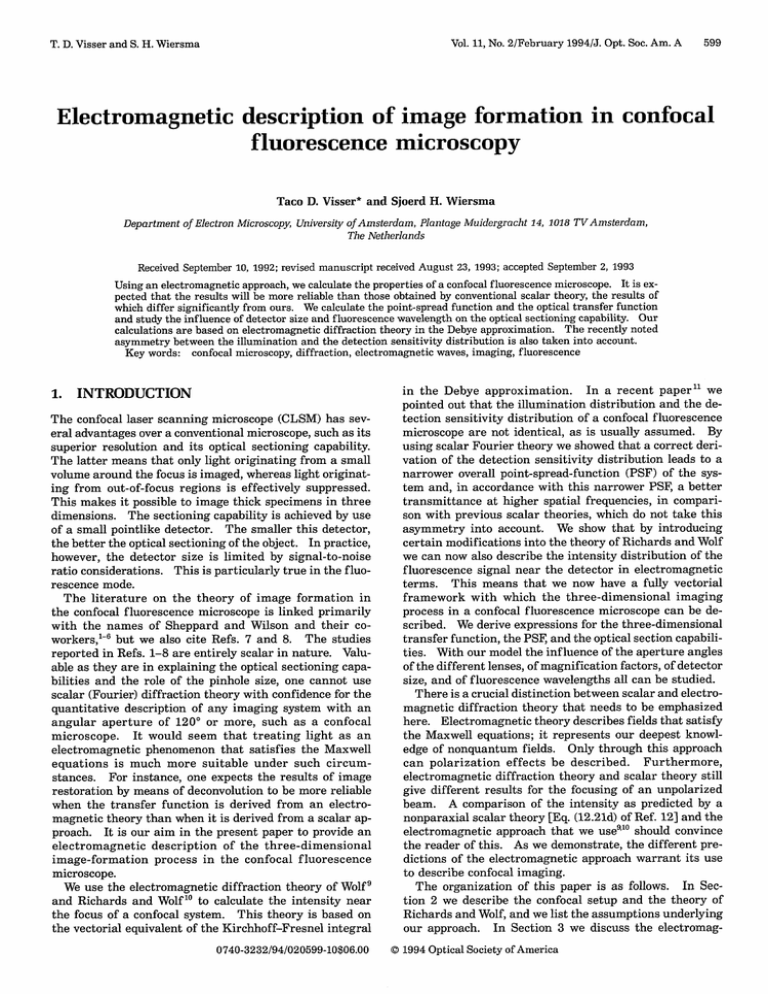
The organization of this paper is as follows. In Section 2 we describe the confocal setup and the theory of
Richards and Wolf,and we list the assumptions underlying
our approach. In Section 3 we discuss the electromag(D1994 Optical Society of America
600
J. Opt. Soc. Am. A/Vol. 11, No. 2/February
T. D. Visser and S. H. Wiersma
1994
detectorplane
collimatedisotropic
fluorescencewave
illuminated in its entirety. That means that f1 2 denotes
the semiangle of the emerging light cone rather than the
semiaperture of the entire lens. Although we restrict
ourselves to an unpolarized incident beam, we need for
purposes later in the paper to define the polarization
angle c. This is the angle between the amplitude Ein, of
the incoming linearly polarized electric field and the positive x axis. The imaging properties of a confocal micro-
scope are strongly determined by the axial intensity
Gaussian
profile
/
expande
dichroic
Gaussian
profilemirror
Fig. 1. Model of the confocal fluorescence
microscope.
originat
An ex-
panded plane laser beam, which is approximately homogeneous, is
focused by Li onto a fluorescent object that can be scanned mechanically. The focus of L, is the origin of the Cartesian coordinates (x, y,z) and the polar coordinates and . L has focal
length fi and semiaperture angle fil. The fluorescent light
originating from the object is collimated by L, and deflected by a
dichroic mirror onto L2 , which in turn focuses the light onto the
pinhole detector. L 2 has focal length f2 and semiaperture angle
f12. The detector, which is placed at the focus of L 2, has a radius
of Vd optical coordinates. The center of the detector coincides
with the center of a second set of coordinates (, 5,i) and i and e.
The wave fronts So, S,, and S2 are discussed in the text.
netic fields on the wave front that are needed for the diffraction integral, for the cases of both illumination and
detection. In Section 4 the intensity in the focal region
and near the detector is calculated. In Section 5 the confocal imaging process is analyzed, and a three-dimensional
optical transfer function is derived. Several generalizations are discussed, as well. In Section 6 the optical
sectioning capabilities of the confocal fluorescence microscope are derived and are compared with results from
scalar theory. The microscope's response to both point
objects (i.e., the PSF) and planar fluorescent objects is
studied for different detector sizes and fluorescence wavelengths. In Section 7 we summarize our results.
2. CONFOCAL MICROSCOPE
A model of the confocal fluorescence microscope is depicted in Fig. 1. The system is symmetric with respect to
rotations around the z and the z axes. A Gaussian beam
with wavelength A. produced by the laser is expanded
such that an effectively uniform plane wave, So,is incident
upon lens Ll with semiaperture angle £l. When no aberrations are present, the wave front after refraction coincides with a reference sphere, or rather with that portion
of it that approximately fills the exit pupil. The reference
sphere, S,, has the focal point as its center and has a radius equal to fi, the focal length of the lens. The focused
light causes an object with a fluorescent material distribution o(x, y, z) to emit fluorescence light. This fluorescence light with wavelength Aftis collected and collimated
by Li and is deflected by the dichroic mirror onto the
second lens, L2 (semiaperture angle f1 2, focal length f2),
which focuses it onto the detection pinhole. The detector
plane coincides with the focal plane of L2. The pinhole
has a radius (expressed in optical coordinates) of Vd In
practice, the radius of L2 may be greater than that of Li,
which means that the aperture of the former will not be
distribution. In a system with rotational symmetry, the
axial intensity distribution is independent of the state of
polarization. In our description we make the basic assumption that the fluorescence response is linear, both in
the fluorescent material density and in the excitation intensity. In Section 5 we discuss possible generalizations
of our approach.
Before analyzing the complete confocal microscope, we
first calculate the electromagnetic field in the focal region
of a high-angular-aperture lens with focal length f and
semiaperture angle Q. In Section 4 we specialize our
results to lenses L, and L 2. Our starting point is the
vectorial theory of Wolf' and Richards and Wolf.'0 This
theory expresses the electric-field amplitude E(x) in the
focal region of a high-aperture lens with focal length f in
terms of the electric field Es in the exit pupil S as
-kf
EW= -1exp(ikf)IEs
I
exp[-ik(Cei
x)]dF,
(1)
with k the wave number and q a unit vector pointing from
the focus in the direction of the point p on the wave front.
The integral is over E;the solid angle subtended by the
aperture at the focal point. Equation (1) is a vector generalization of the Debye integral. 3 It describes the diffracted field as a superposition of plane waves with
amplitudes Es whose propagation vectors all lie within the
geometrical light cone. For an extensive discussion of the
validity of this so-called Debye approximation we refer to
the work of Stamnes.' 2 Here it suffices to say that for a
high Fresnel number system, such as a confocal microscope, the approximation is justified. Equation (1) can
also be obtained if one starts from the Stratton-Chu
integral.' 4 The next ingredient that we need is an expression for the electric-field amplitude Es on the wave front
S for both the illumination and the detection cases. This
will be the subject of Section 3.
3. FIELD AMPLITUDES ON THE WAVE
FRONT
Let Asi denote the amplitude of the electric field on wave
front Si, with (i = 0,1, 2); i.e., ESi = AsiEs, where Es. is a
unit vector. In order to solve Eq. (1) we first consider how
the amplitude As,, of the incident uniform plane wave front
So is connected to the amplitude As8 of the spherical wave
front S, after refraction by lens L, (see Fig. 1). In the
refraction process the energy flux is smeared out. This
projection gives Es, a 0 dependence. Assuming that the
lens obeys the sine condition implies that we are dealing
with the so-called aplanatic energy projection over the
emerging spherical wave front. 2 The sine condition for a
plane wave that is incident parallel to the axis reads as
T. D.Visser and S. H. Wiersma
Vol. 11, No. 2/February
(Ref. 15, p. 168)
(2)
r = f sin 0.
This expression means that the rays enter the lens in the
object space at the same lateral distance r = (X 2 + y 2 )1" 2
from the z axis as the corresponding ray emerges from the
lens in the image space. Furthermore, it implies that a
thin annulus with area S 0 of the incoming plane wave is
projected onto a ring-shaped part of the spherical wave
with area 8S. One can easily show that' 2
aSo = S, Cos ,
0
Conservation of energy then yields for amplitude As, () on
spherical wave front S,
IAsl(0)12 8S,
As o(0)12 8S0 .
=
(4)
For the uniform excitation beam As0 = 1. So the excitation light traveling to focus under an angle 0 with the optical axis has an amplitude
As1(O)= cosl/2
o.
an amplitude Aso(6), for which
.
As(r) = (1
-
-1/4
2
,
r
0
(1
-
M 2 sin 2 6)-1/ 4 Cos1 2 6,
Ai sin
Q1-.
(7)
-
2
-~
~
I0
cs" 6100•42
' '
2
(8)
where we have again used the sine condition but now for
L2 (see also Ref. 17). Notice that for f' = f2 we get
AS 2 (6) = 1. We need not worry that the expression between brackets becomes negative. The lateral coordinate
r has fAsin f 1 as its upper bound. So we have
f2
sin 0 = fi sin 0,
(9)
and hence
(ff
kf,-1
sin2
= sin26
C
0 •C
2,
with the lateral magnification parameter M given by
M =
fi
=
sin f12
(12)
sin f12
Since As1 differs from AS2 we must conclude that the intensity distribution near the focus of Li is not equal to the
distribution near L2. Only in the idealized but physically
unrealistic case of a point source and a point detector3 can
the PSF of a confocal microscope be written as the square
of the excitation intensity. We have shown that when the
finiteness of the source is taken into account, that approximation no longer holds.
4. INTENSITY DISTRIBUTION NEAR
FOCUS AND THE DETECTOR
Suppose that a uniform linearly polarized beam is focused
by a lens. The electric-field amplitude on the converging
spherical wave front can be found by use of geometrical
reasoning. For details we refer to the paper by Richards
and Wolf.'0 An alternative derivation is given by Visser
and Wiersma.'8 It turns out that
Es;a(*,O)= As(0)(E, cos a + E2 sin a),
(10)
which proves the positivity. It is well known that for an
(13)
with
sin
2
4 + cos 0 cos 2
]
E = cos sin 0(cos 0-1)
-sin 0 cos )
cos
At refraction by L2 (notice the change to the second,
tilded [see Eq. (8)] system of coordinates centered at its
focus), the cosine effect of Eq. (5) again occurs. This
means that the emerging converging wave front, which is
focused onto the detector, has an amplitude
AS2(6)= [1 (f) sin2
0
(11)
(6)
Equivalently, one can use the sine condition to express
that the isotropic wave originating from focus after collimation by Li has an amplitude variation
r2
AS 2;M(0) =
(5)
This apodizationlike effect in high-angular-aperture systems was first discussed by Hopkins.'6
Next consider the fluorescence process. The emitted
spherical fluorescence wave has no cos 0 dependence but is
isotropic. When this light, assumed to have unit amplitude, is refracted by Li, the reverse of the above geometrical process takes place. So after collimation the light has
Aso(0) = cos"1/2
601
object in the focal plane of L, that is imaged in the focal
plane of L2, the lateral magnification factor M equals f2/fl.
For the angular dependency of the amplitude on the wave
front S, substituting this gives
(3)
Qfl.
1994/J. Opt. Soc. Am. A
E2 =
,
(14)
J.
(15)
sin (P(cos 0 - 1)
COS2 +
cos 0 sin 2
-sin 0 sin
Here a denotes the polarization angle (the angle between
the incident electric field and the positive x axis), and the
angles and 0 are defined as usual. The amplitude distribution function Asi(0) is defined in Eq. (5) for the focusing of a uniform beam and in Eq. (11) for the case of a
collimated fluorescence beam. Following Richards and
Wolf' 0 we can now solve Eq. (1) for the first lens, Li, with
ES;, given by Eq. (13) andA s l (6) by Eq. (5). The integra-
tion over 4 can be carried out by use of an identity for the
Bessel functions. This can be done for both polarized and
unpolarized light. In the latter case one must also
integrate over the polarization angle a. For the axial intensity (which is the main determinant of the optical sectioning properties) the result, of course, will be identical
for both cases. For a uniform unpolarized incident beam
the outcome for the intensity [or rather the time-averaged
electric energy density (Ref. 15, p. 33)] in the focal region
is
I,,. (q, u)
IIo I ' + 2 I,
12 +
JJ212,
(16)
602
J. Opt. Soc. Am. A/Vol. 11, No. 2/February
T. D. Visser and S. H. Wiersma
1994
Here we used the abbreviation
with
2
Io(u,u) =I Io W,
cos"1
Cos
U)
0
(
iu cos 0 do
sin2 f /(
cos1/2
I,(V ) = |
0
1Afe
sin 0(1 + cos 0)J (~~~~~~sin
snfl,/
sin2
(17)
si
oj
sin 2 Q
9exp(
(25)
Aex
do,
to indicate the ratio of the fluorescence and the excitation
wavelengths. According to Stokes's law we have /3
> 1.
AS;M(0) is given by Eq. (11). Notice that the functions In
depend on v and ii which, just as v and u, are normalized
to l1 and Aexrather than to Q2 and Afl:
(18)
12 (v, u)
J
=
lvsin
cos"/2
0
sin 0(1 - cos 0)J 2 sin
u
N
(19)
here J, is a Bessel function of the first kind, of order n.
[Notice that the angular amplitude distribution function
As(0) is retained in the integrands.] The functions In are
a function of the dimensionless optical coordinates u and
v, which are defined as
v = 7r(X2
Aex
+ y 2)1 2
sin fli,
(20)
Aex
with Aex the excitation wavelength.'9 We also use the coordinates v and v, which are defined in a completely
analogous manner, but then respectively in the x and the y
directions, and for which we have v = (v.2 + vy2)11.
Next we turn to the fluorescence process; that is, we
study how the second lens, L2 , focuses a collimated,
isotropic fluorescence wave, the properties of which were
discussed in Section 3. For the amplitude distribution
function As(0) we must now take Eq. (11). The fluorescence light is assumed to be incoherent and randomly polarized. (Alternatively, one can assume that a fluorescent
point object radiates as the sum of an electric and a magnetic dipole.20 ) Suppose that the detected light has wavelength Afl. Taking into account the different amplitude
distribution function, we now can derive, in the same
fashion as Richards and Wolf did for a uniform wave,' 0 that
for a fluorescence beam the intensity distribution near
the detector at the focus of L2 is, up to a constant, given by
1112 + 211I12 + 11212,
IfI(VU)
(21)
with the functions In defined as
) f A
As
()J+, cOs
M()sn0(
1(
= fl
p sin 2
f
=
-
0 As;M(O)sin
X exp
(26)
In the remainder of the paper we also use O.,and i, for
describing lateral distances in the x and the y directions.
It should be borne in mind that the effective semiaperture angle f12 of the second lens depends on fl and the
total magnification factor M through Eq. (12), from which
f=
sinMi1).
(27)
5. CONFOCAL IMAGING PROCESS
Suppose that we have a point object emitting fluorescent
light with unit intensity, located precisely at the focus of
Li; that is, its position vector in optical coordinates, x, is
given by x = (0, 0, 0). The fluorescent light is collimated
by Li, and after deflection by the dichroic mirror it is focused onto the detector by L2. Hence the intensity in the
detector plane, which we call Id, is now given by Eq. (21);
that is,
Id(, 6y;X) = Ifl(x
Oy, 0),
X
=
(0 0 0).
(28)
Next the point object is shifted laterally to position
x = (vv,,0), while its emission intensity is kept fixed.
The distribution at the detector is then shifted sideways
over a distance (Mvx,Mvy, 0), where M denotes the lateral
linear magnification that is due to the combined action of
Li and L2 [see also Eq. (12)]. Hence
Id(OXvy;x) = IfI(O.- Mv-,
,,
-
Mvy,,0),x = (v,VY, 0).
(29)
For axial displacements the magnification factor is different. According to the lens law, the distance b of the image to a lens and the distance d from the object to the lens
are connected by
b= (
-
1
(30)
(22)
a2 (~~
~sin0
2
As;M(0)sin
fl,
Thus
P in f)
( iacoso
i
2(V)
2)1 2 sin fli.
(£2 +
Aex
sin0
sin
l )
iii os
dp sin 2 fl,
ila )
v=
Akex
X Xepsin
p( iu cos
0)do;
2
f,/
U = 2iZ sin2 1,
sin 2 fy,
=
db
dd
-
(23)
d0
-
0(1-
( ia--sicoso
cos O)J2
-
d i.
3 sin
)
(24)
(1
1 -2 1
b2
f
(31)
So we find that the axial magnification equals, at least
within the focal region, minus the square of the lateral
magnification. That means that the intensity in the detection plane that is due to a fluorescent point object located at x = (v, v, u)is given by
Id(O.,5,,;v.,V",U) = If(O. - MV.,Q- Mvy,,M2 u).
(32)
T. D.Visser and S. H. Wiersma
Now that the system's response to an arbitrarily placed
fluorescent point object (with unit emission intensity) has
been established, we can find the response to an extended
object with a fluorescent material distribution given by
o(x - x,), where x, denotes the scanning position vector
in optical coordinates. We assume that the response of
the fluorescent object is linear in both the excitation intensity lex (i.e., we neglect local saturation effects) and the
density of the fluorescent material. Integration over the
object, with use of Eq. (32), then yields
Id(v
O,
;x3 ) =
ff
X I(.
C(m, n, p)
LLLJ
=
J
J
MVX,
-
2
MV, M u)dvudvydu,
with x = (v.,v,, u) (notice the x, dependence of Id). The
totally measured fluorescence signal F(x,) is equal to the
integral of Id over the detector's surface:
jj
X
If(v6
f
J
Iex(X)O(X
- X.)
- MV,5-
MVYM 2 U)
(34)
X D(63,v5)dvdvydudvkd6,.
Here D(b,, 0) is the detector efficiency function which for
a circular detection pinhole equals
D(6
if 6, 2 + by2
otherwise
by) = {
0
H(vX,,vu; M) =
(3/2
X
(36)
=
ff
-
F(x,) = ffj O(m,n,p)C(m, n,p)
+ nvy, + pu,)]dmdndp,
with the incoherent (or modulation) transfer function
C(m, n,p) defined as
y,
M 2u)
(39)
u)H(vvy,
fIex(vxvy,
M; M)
(40)
Because of the circular symmetry of Iexand H, the transfer function can be written in a partial Hankel form
(Ref. 21, p. 252) as
C(q,p)
=
fJ.
JIex(V,
U)H(V,U; M)
x exp(2wripu)vJo(2rrvq)dvdu,
(41)
with q and p spatial frequencies in the lateral v and the
axial z directions, respectively. From this expression it
follows that the slope of the OTF at q = 0 is zero:
aC(qp)
aq
=
q=0
lim
[
Iex(v,u)H(v, u; M)
qLo fJ Jo
aJ°(2 q) dvdu] .
Sq
(42)
Since lim, _oJo'(x) = 0 one has
aC(q = Op)
0
aq
(43)
For a point detector situated at D. = 6 = 0, which is
represented as D(6 ,v6) = 8(v)8(Q,) the transfer function
takes the simple form
:.i-i'-
C(m,n,p)
=
(37)
If(wx,
2_Y2)12_MvX.
exp[27ri(mv,, + nvy + pu)]dv.dvydu.
Eq. (36) into Eq. (34)
and interchanging the order of integration, we can write
the fluorescence signal as the transform of the product of
O(m, n,p) and the transfer function Cal M,(m, n,p):
) / -Mvx
where we have used the transformation
= 6x - MVX,
and Zy= y - My. The integration is over a circle in the
w&,1~ plane with radius Vd and center (-Mv, -Mvy). Because If, is a circular symmetric function, rotation of the
vector (MvX,Mvy) will alter the domain of integration but
not the outcome. In other words, H is circular symmetric
in its first two arguments, just like I,.. In Appendix A it
is shown that the function H can be transformed into a
single integral. Use of its symmetry in the expression for
the transfer function gives
where the m, n, and p integrations extend from minus in-
X exp[-2wri(mv.,5
-(vd
X exp(2iripu)v
exp[2iri(mvx + nvy + pu)]dmdndp,
Substituting
-
y
2 1 2
d.dwy,
ffj O(m,
n,p)
finity to plus infinity.
2
f(ud -y
-dMVY
X
O(m,
=
U)
From now on we suppress the parameter list (fl , M, 13)in
C(m, n, p), just as overall factors. Next we interchange
the order of integration and first calculate the integral
over the detector. To that purpose, define the function
C(m, n, p)
Vd2
with Vd the detection pinhole radius, expressed in optical
coordinates.
The above derivation of the axial magnification factor
M2 is strictly speaking valid only near the focus, whereas
the three innermost integrations in Eq. (34) are over the
entire R. However, both Ie and Il are sharply peaked
around their respective origins, and hence the main contribution to the integral will come from the focal region.
Next we deduce an expression for the incoherent optical
transfer function (OTF), or modulation transfer function.
To that end one can use the Fourier transform
n,p)
of the fluorescent material density distribution, with m,
n, and p spatial frequencies in the x, y and z (or v.,,vy,u)
directions, respectively, to write
o(x)
2
(38)
fVd-MUY
-
Y-MbY,M
-MV.6,
x D(5,vy)exp[2iri(mv,+ nvy + pu)]dvdvydud3,d5.
(33)
F(xs) = J
IX(X)Il(x
H(vvy,,u;M) as
- x)
ex(x)O(
fl
603
1994/J. Opt. Soc. Am. A
Vol. 11, No. 2/February
x
JIex(X)Ifl(-MV.,-MVy, M2 U)
exp[2iri(mv,,+ mv + pu)]dvdvydu. (44)
(The use of a delta function for a D means that the results
J. Opt. Soc. Am. A/Vol. 11, No. 2/February
604
T. D. Visser and S. H. Wiersma
1994
of scalar theory,4 we see that, although negative values
can also occur here, the electromagnetic approach generally predicts a bandwidth that is filled better (for both a
point and a finite-sized detector). That is, the half-width
of C(q) is a significantly higher percentage of the cutoff
frequency than in the scalar case. All this of course has
consequences for image restoration by deconvolution.
Second, we consider the function C(p) C(q = Op),
the axial variation of the OTF, which is depicted in Fig. 3.
C(p) corresponds to the imaging of thick objects with variations only in the axial direction:
C(p) =
0
0.04
q
->
Fig. 2. (Normalized) lateral variation C(q) of the threedimensional OTF as p = 0 for different radii Vd of the detector.
fl = 600,M = 1OX, = 1.2. In this and subsequent figures the
radii of the pinhole are taken as the size that they have in the
confocal region (i.e., true radius divided by M).
for a finite detector converge to the predictions for a point
detector as d -0.) Using the circular symmetry again
gives
C(qp)
7
I
X
Ie( ,U)fi(MV, M2U)
exp(27ripu)vJo(27rvq)dvdu.
(45)
The incoherent transfer function is a property only of the
microscope itself and not of the object. For any spatialfrequency pair (qp) in the object, the function C(qp)
gives the relative magnitude of the image intensity at
those frequencies. Apart from describing the image formation for an arbitrary object, the OTF is also a useful
tool for image restoration. (See also the remarks at the
end of this section.) For further background on the OTF
in confocal microscopy we refer to the paper by Sheppard
and Gu.22
From Eq. (45) it can be seen that the transfer function
for a point detector system is the transform of the product
of the excitation intensity and the intensity at the detector that is due to a fluorescent point object. Notice that
this interpretation is independent of the precise form of
the two distributions. We consider next two special cases
of Eq. (41). First we consider the function C(q)C(q,p = 0), which describes the imaging of thick objects
with variations only in the lateral direction:
C(q) =
f
vJo(27rvq)[
f
f Iex(, u)H(v, u; M)v cos[27rpu]dvdu.
(47)
0.08
Iex(v, u)H(v, u; M)du] dv.
Here we see a decrease of the OTF with increasing pinhole
size. The deviations from the scalar theory are similar to
those of the lateral case. If we normalize p and q to
sin2 fl, and sin fli, respectively [see Eqs. (20)], it is found
(for the choice of parameters in Figs. 2 and 3) that the
resolution in the lateral direction is approximately three
times better than the axial resolution.
As an aside, we point out that three generalizations can
easily be incorporated into our framework. First, one can
assume the incident light to be linearly polarized. This
would simply mean that an integration over all polarization angles is left out of the theory (see Ref. 10). The intensity near focus, and hence the response of the system,
is then of course no longer axially symmetric.23 Second,
one may want to take aberrations into account by letting
the diffraction integral extend over the deformed wave
front. This idea is worked out by Visser and Wiersma.18
Third, the effect of different beam profiles, such as centrally obscured beams and Gaussian beams (or, equivalently, apodized lenses) on the focal electromagnetic-field
distribution can be studied by use of the results described
in Ref. 14.
Another matter is the influence of the object that is imaged. This influence can take several forms. Scattering
and absorption of both the excitation and the fluorescence
light may cause the contribution of points deep within
the object to become attenuated. An image-processing
method has been proposed to compensate for this effect.2 4
(46)
Because of symmetry the integral over u extends over '
only. In Fig. 2, C(q) is shown for several detection pinhole sizes. The transfer function narrows with increasing pinhole radius Vd. Also, the cutoff frequency strongly
decreases. For pinhole radii greater than 2.5 the OTF
can actually become negative, with the tail of the transfer
function oscillating. The negative values of the OTF
cause contrast reversals for fine details in the object and
hence cause the image fidelity to deteriorate. (Note that
the OTF for a single aberration-free lens cannot become
negative.) If we compare Eq. (46) with the predictions
0
0.05
0.10
0.15
Fig. 3. (Normalized) axial variation C(p) of the threedimensional OTF as q = 0 for different radii ud of the detector.
fl = 60',M = 10X, P = 1.2. The curves for a point detector
and Vd = 2.5 are very close together on the scale of this figure.
Vol. 11, No. 2/February
T. D. Visser and S. H. Wiersnia
1994/J. Opt. Soc. Am. A
605
detection pinhole sizes. (The numerical integrations
were carried out with routine DO1AKF of the NAG library.27 ) It is clear that a larger pinhole size leads to an
increased half-width of the function F(u). In other words,
the sectioning decreases when the pinhole becomes larger.
On the scale of this figure the curves for a true point detector and one with a radius Vd = 2.5 optical units can
hardly be distinguished. So one can increase the pinhole
diameter to a certain limit in order to improve the signalto-noise ratio without causing the optical sectioning to deteriorate. On the other hand, it also follows from Fig. 4
that for large pinhole sizes the curves for F(u) change less
and less with increasing radius until the entire diffraction
10
D
pattern is picked up by the detector. Any further increment of the detector size will result only in a decrease of
uo
the signal-to-noise ratio. Equation (50) can also be used
Fig. 4. Detected signal for a point object, axially scanned
through focus, for different pinhole radii d. The sem:iaperture
to study the axial resolution of the microscope. Because
angle of the illumination lens Li is 60°, the magnifical tion
M is
we are dealing with a linear system, we can find the relOx, and 3 = Afi/Aex= 1.2. The response is normalize
d toF( sponse to two point objects a distance Auapart on the axis
A refractive-index mismatch between the (oil) im mersion
fluid and the (watery) object leads to two effects First,2 5
dati*n
there is a spherical aberrationlike image degra( catonb
Second, and even more important, the true dist. 3.nce etween the optical sections is then no longer equEil to the
distance over which the object stage has been mcwved. If
this effect is not taken into account, as is freque ntly the
case, objects may appear to be as much as thrEwe times
larger than they actually are.26
6. OPTICAL SECTIONING
As noted above, the optical sectioning capability of a confocal fluorescence microscope is achieved by use o:f a small
pinhole in front of the detector. This sectioning ]property
of a CLSM can be described by its response to a n object
that is axially scanned through the focus. In otheor words,
we want to know how the measured intensity del)ends on
the position of the object that is imaged. Two oither parameters that we study below are the pinhole size and the
fluorescence wavelength. We therefore return to Eq. (34).
For the sake of brevity we omit the scanning po 3ition x5
from now on. Two suitable objects for our purpc)se are a
fluorescent point object and an infinitesimally thin plane
with a uniform fluorescence distribution. First, consider
the former case: a fluorescent point object pIlaced at
(0,0, u'). The object function o is then given by
o(x) = (v)8(v)5(u -
').
by evaluating F(u) and F(u + Au) and adding the results.
This is shown in Fig. 5 for several values of Au. When
au = 5.0 axial optical units (not shown), the width of the
response is increased by 42% compared with the curve for
a single point, but the curve still has a single peak. In
other words, one cannot really resolve the two points. For
6u = 7.5 the dip is 93% of the peak value.
When Au is
further increased to 10.0, the dip reduces to a mere 52%.
The second object that we study is an infinitely thin
plane with a uniform surface distribution of fluorescent
material. The plane is perpendicular to the u axis and is
located at u = u'. We then have
O(x)= (u - ').
(51)
Substituting this into Eq. (34) gives
_".
F(u')
f_.
f~M
J
J
=
J
2
(Id
2
-
112
Y )
- VdxMVy F(Id2_6Y2)1/2_
-M')
MI.
f (x,&Y, M2U ')d&dwydv.,dvy,
x
(52)
where we have used the transformation
, = 6 - MVX
and &y= y - Mvy. In this expression we recognize the
F(u)
(48)
Using that I(0,u) = I2(0,u) = I1(0,7)= I2 (0,ir)= 0 for
all u and ii and exploiting the rotational symmetry yield
F(6;O.O. ') = I( , 0 U) 2 " I ( M2U,)Off
(49)
~~ ~~~~~~~~=1
0~
So now we have found the axial PSF for the CLSM. For a
point detector at v = 0, Eq. (34) reduces to
F(O,0,u') = IIo(0,0, u')Io(0, M2 u')2.
(50)
Notice that, because the optical coordinate v is dimensionless, F has the same dimension in Eqs. (49) and (50). In
Fig. 4 we plot the detected signal F as a function of the
axial position u of a fluorescent point object for different
0
5
10
15
20
25
30
35
40
Up
Fig. 5. Response of a confocal fluorescence microscope with a
point detector for two point objects on the axis that are a distance
Su apart.
All curves are normalized to unity.
l = 60,
M = OX,,G= 1.2. For clarity the curves have been displaced
with respect to one another.
606
J. Opt. Soc. Am. A/Vol. 11, No. 2/February
T. D. Visser and S. H. Wiersma
1994
F(u)
0
2
4
6
8
U p
10
Fig. 6. Detected signal for a perfect planar fluorescent object,
axially scanned through the focus of a confocal fluorescence
microscope for different values of the detection pinhole radius ud.
Q1 = 600,M = lox,3 = 1.2.
previously defined function H(v, u; M) of Eq. (39). Substitution yields
FW) = fo, I,.(v, u')H(v, u')vdv.
tern at the detector becomes very sensitive to the extent
to which lens L2 is filled (depending of course on the
distance L,-L2), causing the axial response of the microscope to become asymmetrical. This last point seems
clearly an undesired feature of such a design. Incidentially, a vectorial theory that is valid for both high and
very low angular aperture focusing has recently been
formulated. 4
The next parameter of interest is G3= AfI/Aex,
the ratio
of the excitation and the fluorescence wavelengths. In
Fig. 7 the response of a system with a point detector to an
axially scanned point object is depicted for several values
of G3. The limiting value j3 = 1 gives the optimal result.
From the figure it followsthat it is best to keep Aflas close
to Aexas possible. The dashed curve is the prediction of
scalar theory 3 for /3 = 1.0,which differs significantly from
the outcome of our model.
In Fig. 8 the response of a point detector CLSM to an
axially scanned fluorescent planar object is shown for different values of /3. If we compare these results with the
predictions of scalar theory 2 we find a strong deviation.
(53)
For the case of a point detector at v = 0, we have
and the expression for F(u')becomes
D(Dx,6,) = 8(ix)8(,)
F(u') =
f
Iex(V,u')If(-MV, M2u')vdv.
(54)
The sectioning of a plane is worse than that of a point
object, as follows from a comparison of Fig. 4 with Fig. 6.
For instance, the half-width of F(u) when a plane is
imaged with Vd= 5.0 and 3 = 1.2 is approximately 35%
greater than when a point is imaged under the same circumstances. The fact that the sectioning of a planar object is worse than that of a point can be understood by
consideration of the intensity contours in the v, u plane,2 3
from which it can be seen that the intensity I.. (and hence
also in approximation the function if,)for off-axis points
within the central peak decreases much less than for a
point on the axis when a small excursion in the u direction
is made. It should also be noted that for very low aperture angles (i.e., fl s 10°) we retrieve the results from
classical paraxial scalar theory.2 Also, as can be seen
from Fig. 6, the sectioning of a plane is much more sensitive to an increase in the pinhole size than that of a single
point. This is because the diffraction pattern at the detector extends over a larger area for a plane than for a
point object.
The lateral magnification factor M enters our equations
in two ways: first, in the argument of the function If, [as
in Eq. (49)], and second, through the M dependence of the
semiaperture angle f1 2 [see Eq. (27)]. For values of M
between 5 and 1000 the curves in Figs. 4 and 6 remain
practically identical, as one might expect. For M between
1 and 5 however, deviations up to 15% in the values of F(u)
occur. When M gets larger than -1000, the semiaperture
fi2 of the second lens L2 becomes so small that the Debye
approximation, and hence our diffraction integral, is no
longer valid.28 For such lenses the so-called focal shift
phenomenon occurs.
This means that the intensity pat-
u £
Fig. 7. Response of a confocal fluorescence microscope with a
point detector imaging a point object that is axially scanned
through focus for different values of ,3= Afl/Aex. Notice the difference between the predictions of electromagnetic theory (solid
curves) and of scalar theory (dashed curve).
l, = 600 and
M = lox.
0
2
4
6
8
U p>
10
Fig. 8. Detected signal for a perfect planar fluorescent object,
axially scanned through the focus of a confocal fluorescence microscope with a point detector for different values of : = Af1/Aex.
flI = 60° and M = 10x. Dashed curve, the prediction of scalar
theory; solid curves, the results according to electromagnetic diffraction theory. Note the large difference between them.
Vol. 11, No. 2/February
T. D. Visser and S. H. Wiersma
607
1994/J. Opt. Soc. Am. A
two-plane resolution is worse than the resolution for a twopoint object.
F(u)
7.
CONCLUSION
Wehave presented an electromagnetic theory of the confomicroscope.
cal fluorescence
-
0
0
20
10
6u=1O~
~
40
30
u =15.0
50
60
70
u
s>
Fig. 9. Response of the confocal fluorescence microscope's imag-
ing of two planes, both of which are perpendicular to the central
axis. The distance between the planes is Su optical coordinates.
For clarity the curves have been displaced with respect to each
other.
Not only do our results dif-
fer significantly from scalar theory, as we have shown with
several examples, but we also expect that our approach
will give a much more precise description of the imaging
process than any scalar theory would. The influence of
several factors such as magnification, detection pinhole
size, and fluorescence wavelength was studied. It was
found in our approach that the modulation transfer function can become negative (even for an aberration-free system) when the detection pinhole size exceeds a certain
value. This transfer function can be used for image restoration by deconvolution of confocal images and should
give more reliable results than those derived from scalar
theory. We calculated the (axial) point-spread function
and showed that the optical sectioning of a plane is worse
than that of a point object. Also, the former is more
sensitive to imaging parameters such as detection pinhole
size and fluorescence wavelength. Finally, our analysis
showed that when the finiteness of the laser source is
taken into account, the overall point-spread function is no
longer the square of the excitation intensity.
FUNCTION H(v, u; M)
APPENDIX A:
In this appendix we evaluate the function H(v,v,
as a single line integral. From Eq. (39) we have
u; M)
Vd 2 Mv
(a)
H(vx,vy, u; M)
-1
Wy
'My
_ d-MVY
J(vd
f(vd
-
-y2)
M.
ifi(&., &Y,M 2 u)d&,,d&y. (Al)
2
-uY2)112-mv,
The integration in the wiiZy plane is over a circle with
center (-Mv,, -Mvy) and radius Vd. Because of the
circular symmetry, we have H(v, 0, u';M) = H(v,, vy,u'; M),
with v = ( 2 + vY2 )1/2. The left-hand side is easier to
analyze. First, consider the case in which Vd - MV
[Fig. 10(a)]. Using polar coordinates, we transform H into
T
CMV+vd fo(r)
H(v, 0, u;
I
M)d2MV
=
J
Vd <MV
M +Vd
(b)
= fJ
Fig. 10. Region of integration for the function H(v, u; M) for the
case in which (a) d - Mv and (b) Vd < Mu
For /3= 1.0, scalar theory predicts a better sectioning.
In addition to the inferior sectioning of a plane compared
with that of a point and the greater sensitivity of a plane
to the pinhole size, the sectioning of a plane deteriorates
much more strongly than the sectioning of a point object
when /3gets larger.
Finally, the axial two-plane resolution of the confocal
microscope is depicted in Fig. 9, where the response to two
parallel planes perpendicular to the u axis is shown for two
values of Au,their mutual distance. Contrary to the case
in Fig. 5, the curve for Au = 7.5 (not shown) is now still a
single peak. For Su = 10.0 the dip is higher than for the
two-point case. This is another way of showing that the
Ifl(r)rd4)dr
(A2)
2If1(r)r(D(r)dr,
(A3)
f
with ¢(r) now to be determined. As can be seen from the
figure, the 0 integration is over the entire 27ras long as
0 ' r ' Vd - Mu For larger values of r the integration
is limited to the hatched circle segment, which is bounded
by the intersection of the curves &j,2 + y2= r2 and
(lb" - Mv)2 + Wy2= Vd2. From the above it follows that
Wx=
Vd 2-r2
M2V2
So the point of intersection makes an angle
positive &, axis, for which
4Vd=2r2 - M2v2)
d = os'V)
(A4)
-2Mv
4 with
the
(A5)
608
J. Opt. Soc. Am. A/Vol. 11, No. 2/February
Summarizing, we have for
Vd -
2 - r2
cos-'[(Vd
8. S. Kawata, R. Arimoto, and 0. Nakamura, "Three-
Mv that
-
r
d-MV
M 2 v2 )/-2Mvr]
dimensional optical-transfer function analysis for a laserscan fluorescence microscope with an extended detector,"
if 0
Ir
lD(r)dmV=
T. D. Visser and S. H. Wiersma
1994
* (A6)
otherwise
10. B. Richards and E. Wolf, "Electromagnetic
The other case that we need to consider is that in which
Vd < Mu We then have from Fig. 10(b)
f-Mv+vdfo(r)
H(v,0, u;M)vd<Mv =
J
J
rMV+Vd
=
I
M -ud
J. Opt. Soc. Am A 8, 171-175 (1991).
9. E. Wolf, "Electromagnetic diffraction in optical systems I,"
Proc. R. Soc. London Ser. A 253, 349-357 (1959).
If (r)rdodr
1
(A7)
diffraction
in op-
tical systems II," Proc. R. Soc. London Ser. A 253, 358-379
(1959).
11. T. D. Visser, G. J. Brakenhoff, and F. C. A. Groen, "The
fluorescence point response in confocal microscopy," Optik
87, 39-40 (1991).
12. J. J. Stamnes, Waves in Focal Regions (Hilger, Bristol, UK,
1986).
Vr
2If(r)r(D(r)dr.
(A8)
13. P. J. W Debye, "Das Verhalten von Lichtwellen in der Nihe
eines Brennpunktes oder einer Brennlinie," Ann. Phys. 30,
755-776 (1909).
14. T. D. Visser and S. H. Wiersma, "Diffraction of converging
electromagnetic beams," J. Opt. Soc. Am. A 9, 2034-2047
In precisely the same manner as above we now get
(1992).
15. M. Born and E. Wolf, Principles of Optics, 6th ed. (Perga(r)Vd<MV = Cos 1(
)2 *
(A9)
This concludes the transformation of H into a single line
integral.
ACKNOWLEDGMENTS
mon, Oxford, UK, 1980).
16. H. H. Hopkins, "The Airy disc formula for systems of high
relative aperture," Proc. Phys. Soc. 55, 116-128 (1943).
17. R. Barakat and D. Lev, "Transfer functions and total illuminance of high numerical aperture systems obeying the sine
condition," J. Opt. Soc. Am. A 53, 324-332 (1963).
18. T. D. Visser and S. H. Wiersma, "Spherical aberration and the
electromagnetic field in high-aperture systems," J. Opt. Soc.
Am. A 8, 1404-1410 (1991).
*Present address, Department of Physics and Astronomy, Free University, De Boelelaan 1081, 1081 HV Amsterdam, The Netherlands.
19. An alternative definition is given by Sheppard and Matthews
[J. Opt. Soc. Am. A 4, 1354-1360 (1987)] that has the advantage that the axial intensity distribution is less sensitive to
the aperture angle. Here, however, we follow the definition
given by Richards and Wolf.
20. C. J. R. Sheppard and T. Wilson, "The image of a single point
in microscopes of large numerical aperture," Proc. R. Soc.
London Ser. A 379, 145-158 (1982).
21. R. N. Bracewell, The Fourier Transform and Its Applications
REFERENCES AND NOTES
22. C. J. R. Sheppard and M. Gu, "The significance of 3D transfer
functions in confocal scanning microscopy," J. Microsc. 165,
We thank Koen Visscher for many discussions on the confocal microscope. We also thank H. A. Ferwerda and
Colin Sheppard, who suggested several improvements in
the original manuscript.
(McGraw-Hill, New York, 1986).
1. C. J. R. Sheppard and T. Wilson, "Image formation in scanning microscopes with partially coherent source and detector," Opt. Acta 25, 315-325 (1978).
2. T. Wilson, "Optical sectioning in confocal fluorescent microscopes," J. Micros. 154, 143-156 (1989).
3. T. Wilson and C. J. R. Sheppard, Theory and Practice of
Scanning Optical Microscopy (Academic,London, 1984).
4. M. Gu and C. J. R. Sheppard, "Confocal fluorescent microscopy with a finite-sized circular detector," J. Opt. Soc.Am. A
9, 151-153 (1992).
5. T. Wilson, "The role of the pinhole in confocal imaging systems," in Handbook of Biological Confocal Microscopy, J. B.
Pawley, ed. (Plenum, New York, 1990).
6. C. J. R. Sheppard, 'Axial resolution of confocal fluorescence
microscopy," J. Microsc. 154, 237-241 (1989).
7. S. Kimura and C. Munakata, "Calculation of threedimensional optical transfer function for a confocal scanning
fluorescent microscope," J. Opt. Soc. Am. A 6, 1015-1019
(1989).
377-390 (1992).
23. A. Boivin and E. Wolf, "Electromagnetic field in the neighborhood of the focus of a coherent beam," Phys. Rev. 138,
B1561-B1565 (1965).
24. T. D. Visser, F. C. A. Groen, and G. J. Brakenhoff,
'Absorp-
tion and scattering correction in fluorescence confocal microscopy," J. Microsc. 163, 189-200 (1991).
25. C. J. R. Sheppard and C. Cogswell, "Effect of aberrating
lay-
ers and tube length on confocal imaging properties," Optik
87, 34-38 (1991).
26. T. D. Visser, J. L. Oud, and G. J. Brakenhoff, "Refractive
index and distance measurements in 3-D microscopy," Optik
90, 17-19 (1992).
27. NumericalAlgorithm GroupFORTRAN
Library Manual,Mark
14 (NAG Ltd., Oxford, UK, 1991).
28. E. Wolf and Y Li, "Conditions for the validity of the Debye
integral representation of focused fields," Opt. Commun. 39,
205-210 (1981).
29. Y. Li and E. Wolf,"Focal shift in focused truncated Gaussian
beams," Opt. Commun. 42, 151-156 (1982).


