Emergency - Tachyarrhythmias
advertisement
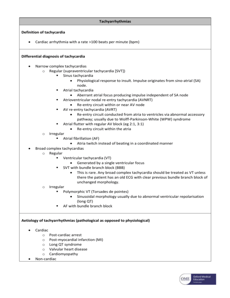
Tachyarrhythmias Definition of tachycardia Cardiac arrhythmia with a rate >100 beats per minute (bpm) Differential diagnosis of tachycardia Narrow complex tachycardias o Regular (supraventricular tachycardia [SVT]) Sinus tachycardia Physiological response to insult. Impulse originates from sino-atrial (SA) node. Atrial tachycardia Aberrant atrial focus producing impulse independent of SA node Atrioventricular nodal re-entry tachycardia (AVNRT) Re-entry circuit within or near AV node AV re-entry tachycardia (AVRT) Re-entry circuit conducted from atria to ventricles via abnormal accessory pathway; usually due to Wolff-Parkinson-White (WPW) syndrome Atrial flutter with regular AV block (eg 2:1, 3:1) Re-entry circuit within the atria o Irregular Atrial fibrillation (AF) Atria twitch instead of beating in a coordinated manner Broad complex tachycardias o Regular Ventricular tachycardia (VT) Generated by a single ventricular focus SVT with bundle branch block (BBB) This is rare. Any broad complex tachycardia should be treated as VT unless there the patient has an old ECG with clear previous bundle branch block of unchanged morphology. o Irregular Polymorphic VT (Torsades de pointes) Sinusoidal morphology usually due to abnormal ventricular repolarisation (long QT) AF with bundle branch block Aetiology of tachyarrhythmias (pathological as opposed to physiological) Cardiac o Post-cardiac arrest o Post-myocardial infarction (MI) o Long QT syndrome o Valvular heart disease o Cardiomyopathy Non-cardiac o o o Hypoxia Hypovolaemia Electrolyte abnormalities Especially hypo/hyper-kalaemia, -calcaemia or -magnesaemia o Hypoglycaemia o Hypo/hyperthermia o Hypo/hyperthyroidism o Sepsis Drug-induced o Cocaine o Amphetamines o Tricyclic antidepressants o Beta blockers o Digoxin o Amiodarone Clinical features of tachycardias Adverse features o Shock Hypotension, diaphoresis, pallor, increased capillary refill time (CRT) o Syncope Transient loss of consciousness o Myocardial ischaemia Ischaemic chest pain and/or ischaemic electrocardiogram (ECG) changes o Cardiac failure Orthopnoea, paroxysmal nocturnal dyspnoea (PND), bibasal crepitations, raised jugular venous pressure (JVP) Non-adverse features o Palpitations o Dyspnoea o Anxiety Initial investigation of tachycardia Bloods o Full blood count o Urea & electrolytes o Magnesium o Bone profile (particularly noting calcium and phosphate) o Thyroid function tests o Other: liver function (useful pre-medication); coagulation (may need anticoagulation) Chest radiograph (CXR) Further investigation of tachycardia Echocardiogram (echo) Initial management of tachycardia Assess patient from an ABCDE perspective Maintain a patent airway o Use manoeuvres, adjuncts, supraglottic or definitive airways as indicated Controlled oxygen o Maintain saturations (SpO2) 94-98% Attach monitoring o Pulse oximetry o Non-invasive blood pressure o Three-lead cardiac monitoring o Defibrillator pads 12 lead ECG Obtain intravenous (IV) access and take bloods Give IV fluid challenge if appropriate and repeat as necessary Identify and treat any reversible causes e.g. electrolyte abnormalities on initial VBG If adverse features are present [shock, syncope, myocardial ischaemia, heart failure] prepare for DC cardioversion under general anaesthesia or conscious sedation o Once ready, warn all those nearby to stand clear and remove any oxygen delivery device whilst the defibrillator is set to synchronised mode and charged to 120 J o Once the defibrillator is charged and all are clear, deliver the shock o Should this fail, two subsequent shocks at increasing increments may be tried o Should this fail, give a loading dose of amiodarone 300 mg IV over 10-20 minutes and repeat DC cardioversion followed by amiodarone 900 mg IV over 24 hours If adverse features are not present, assess the rhythm: Narrow complex tachycardias (QRS duration <0.12 s) o Regular: likely SVT Attempt vagal manoeuvres Valsalva (ask patient to blow into syringe); carotid sinus massage. o If this fails then: Adenosine 6 mg IV Rapid bolus ideally into a large-bore cannula in the antecubital fossa Warn patients of transient unpleasant side effects: flushing, nausea and chest tightness, ‘feeling of impending doom’ Avoid in patients with asthma, WPW syndrome, and denervated hearts Caution in taking throphylline, dipyridamole or carbamazepine If 6mg unsuccessful: Adenosine 12 mg IV If first 12mg unsuccessful: Further adenosine 12 mg IV If adenosine is contraindicated, consider verapamil 2.5-5.0 mg IV, or flecainide 2 mg/kg IVI over 20-30 min if no evidence of structural heart disease o Irregular: likely AF Onset <48 hours Aim for rhythm control o Flecainide 2 mg/kg IVI over 20-30 min if no evidence of structural heart disease or amiodarone 300 mg IV over 20-30 min and 900 mg over 24 hours if flecainide contraindicated o Anticoagulate with enoxaparin 1.5 mg/kg subcutaneous (SC) prior to this Onset >48 hours Aim for rate control o Metoprolol 5 mg IV OR bisoprolol 5 mg orally (PO) OR verapamil 5 mg IV If signs of heart failure try digoxin 0.5 mg IVI over 30-60 min Digoxin can be added to the above if beta-blockade unsuccessful o Anticoagulate with enoxaparin 1.5 mg/kg subcutaneous (SC) prior to this Broad complex tachycardias (QRS duration >0.12 s) o Regular If likely monomorphic VT Give amiodarone 300 mg IVI over 20-30 min followed by amiodarone 900 mg IVI over 24 hours Any broad complex tachycardia should be treated as VT unless there the patient has an old ECG with clear previous bundle branch block of unchanged morphology. If definitely SVT with BBB Try adenosine as for regular narrow complex tachycardias o Irregular If likely AF with BBB Treat as for irregular narrow complex tachycardias If likely polymorphic VT (Torsades de pointes) Magnesium 2 g IV over 10 min Stop any medications which prolong the QT interval Correct any electrolyte abnormalities if not already done so, and give Further management of tachycardia Request 12 lead ECG once back in sinus rhythm o Look specifically for ischaemic changes (ST elevation, ST depression and T wave inversion), prolonged QT interval (QTc >440 ms) and signs of WPW syndrome (shortened PR interval, delta wave and broad QRS complex) Identify and correct any underlying cause if not already done so Call cardiologist o Arrange for an implantable cardioverter defibrillator (ICD) if appropriate Common questions concerning tachycardia List the adverse features which signify an unstable tachyarrhythmia What is the appropriate management of a tachyarrhythmia if one or more adverse features are present? What procedure must be performed in a conscious patient in order to facilitate this management? If no adverse features are present, outline your approach to the assessment of a stable tachyarrhythmia How would you manage a patient in stable SVT? How would you manage a patient in stable AF of onset <48 hours? How would you manage a patient in stable AF of onset >48 hours? How would you manage a patient in stable VT? Once the tachyarrhythmia has reverted back to sinus rhythm, what follow up investigations would you request?
