(18) Urinary System Anatomy
advertisement
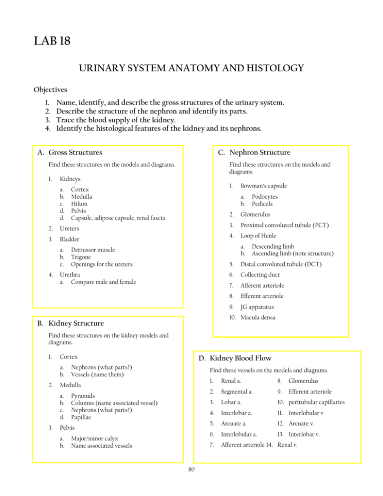
LAB 18 URINARY SYSTEM ANATOMY AND HISTOLOGY Objectives 1. 2. 3. 4. Name, identify, and describe the gross structures of the urinary system. Describe the structure of the nephron and identify its parts. Trace the blood supply of the kidney. Identify the histological features of the kidney and its nephrons. A. Gross Structures C. Nephron Structure Find these structures on the models and diagrams. 1. Kidneys a. b. c. d. d. Ureters 3. Bladder a. b. c. 1. Cortex Medulla Hilum Pelvis Capsule, adipose capsule, renal fascia 2. 4. Find these structures on the models and diagrams. Bowman’s capsule a. b. 2. Glomerulus 3. Proximal convoluted tubule (PCT) 4. Loop of Henle a. b. Detrussor muscle Trigone Openings for the ureters Urethra a. Compare male and female Podocytes Pedicels Descending limb Ascending limb (note structure) 5. Distal convoluted tubule (DCT) 6. Collecting duct 7. Afferent arteriole 8. Efferent arteriole 9. JG apparatus 10. Macula densa B. Kidney Structure Find these structures on the kidney models and diagrams. 1. Cortex a. b. 2. Find these vessels on the models and diagrams. Medulla a. b. c. d. 3. D. Kidney Blood Flow Nephrons (what parts?) Vessels (name them) Pyramids Columns (name associated vessel) Nephrons (what parts?) Papillae Pelvis a. b. Major/minor calyx Name associated vessels 80 1. Renal a. 8. Glomerulus 2. Segmental a. 9. Efferent arteriole 3. Lobar a. 10. peritubular capillaries 4. Interlobar a. 11. Interlobular v 5. Arcuate a. 12. Arcuate v. 6. Interlobular a. 13. Interlobar v. 7. Afferent arteriole 14. Renal v. E. Nephron Histology Find these structures on the slide of the kidney and any model that pertains. Describe the histological characteristics of each structure and define its function. 1. Cortex 2. Medulla 3. Renal corpuscle 4. Bowman’s capsule 5. Glomerulus 6. Proximal convoluted tubule 7. Distal convoluted tubule 8. Collecting duct F. Kidney Dissection Obtain a sheep kidney. 1. Locate and identify external structures before cutting 2. Identify if it is a right of left kidney 3. Remove the fascia (if present) and identify the capsules 4. Cut into sagittal sections and identify any visible internal structures G. Questions 1. Define retroperitoneal. 2. Which renal artery is longest? Why? 3. Name two types of nephrons. 4. What parts of the nephron are associated with the cortex? 5. What parts of the nephron are associated with the medulla? 6. Trace the blood flow through the kidney. 81 7. What is the anatomical position of the kidneys? 8. Distinguish between internal and external urethral sphincters. 9. What is the detrussor muscle? 10. What is the Juxtaglomerular apparatus and where is it located? Distinguish between the JG cells (location and function) and the macula densa cells (location and function). 11. Give the location and function of the peritubular capillaries. H. Label the structures on the diagrams below 1 2 (region) (covering) 3 (region) 11 4 10 5 9 8 6 7 82 12 17 (entire structure) 13 15 14 16 18 19 20 21 22 24 23 83
