abstract - Smart Materials and Sensors
advertisement
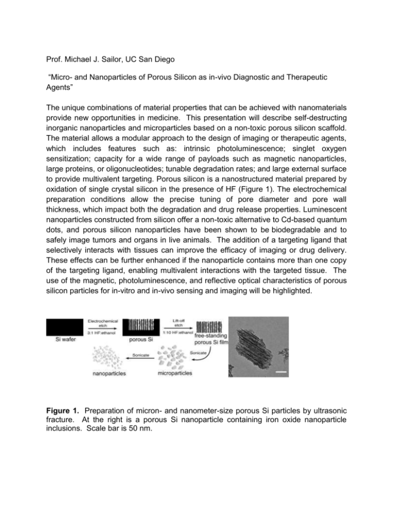
Prof. Michael J. Sailor, UC San Diego “Micro- and Nanoparticles of Porous Silicon as in-vivo Diagnostic and Therapeutic Agents” The unique combinations of material properties that can be achieved with nanomaterials provide new opportunities in medicine. This presentation will describe self-destructing inorganic nanoparticles and microparticles based on a non-toxic porous silicon scaffold. The material allows a modular approach to the design of imaging or therapeutic agents, which includes features such as: intrinsic photoluminescence; singlet oxygen sensitization; capacity for a wide range of payloads such as magnetic nanoparticles, large proteins, or oligonucleotides; tunable degradation rates; and large external surface to provide multivalent targeting. Porous silicon is a nanostructured material prepared by oxidation of single crystal silicon in the presence of HF (Figure 1). The electrochemical preparation conditions allow the precise tuning of pore diameter and pore wall thickness, which impact both the degradation and drug release properties. Luminescent nanoparticles constructed from silicon offer a non-toxic alternative to Cd-based quantum dots, and porous silicon nanoparticles have been shown to be biodegradable and to safely image tumors and organs in live animals. The addition of a targeting ligand that selectively interacts with tissues can improve the efficacy of imaging or drug delivery. These effects can be further enhanced if the nanoparticle contains more than one copy of the targeting ligand, enabling multivalent interactions with the targeted tissue. The use of the magnetic, photoluminescence, and reflective optical characteristics of porous silicon particles for in-vitro and in-vivo sensing and imaging will be highlighted. Figure 1. Preparation of micron- and nanometer-size porous Si particles by ultrasonic fracture. At the right is a porous Si nanoparticle containing iron oxide nanoparticle inclusions. Scale bar is 50 nm.
