Supplementary data Fig. 1: Darkfield image and AFM image of 80
advertisement
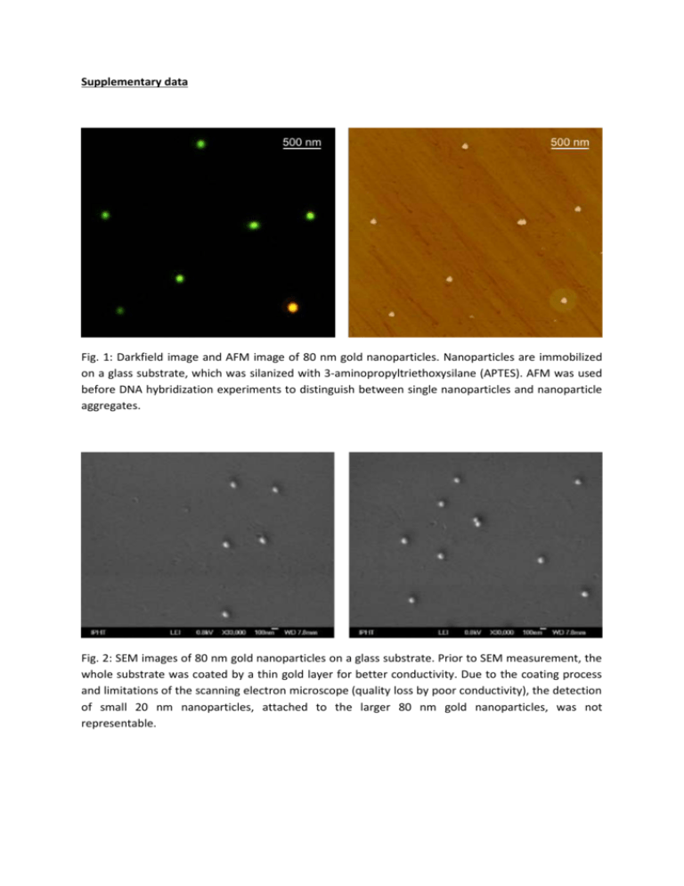
Supplementary data Fig. 1: Darkfield image and AFM image of 80 nm gold nanoparticles. Nanoparticles are immobilized on a glass substrate, which was silanized with 3-aminopropyltriethoxysilane (APTES). AFM was used before DNA hybridization experiments to distinguish between single nanoparticles and nanoparticle aggregates. Fig. 2: SEM images of 80 nm gold nanoparticles on a glass substrate. Prior to SEM measurement, the whole substrate was coated by a thin gold layer for better conductivity. Due to the coating process and limitations of the scanning electron microscope (quality loss by poor conductivity), the detection of small 20 nm nanoparticles, attached to the larger 80 nm gold nanoparticles, was not representable.


