Micro-Viewer Lab: Animal Mitosis NAME: Introduction: Throughout
advertisement
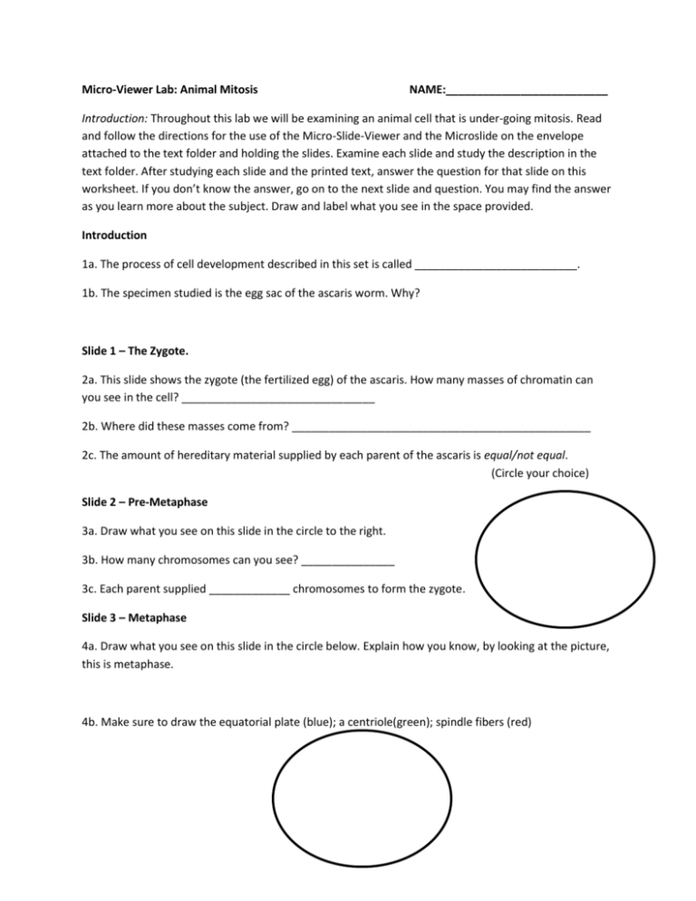
Micro-Viewer Lab: Animal Mitosis NAME:__________________________ Introduction: Throughout this lab we will be examining an animal cell that is under-going mitosis. Read and follow the directions for the use of the Micro-Slide-Viewer and the Microslide on the envelope attached to the text folder and holding the slides. Examine each slide and study the description in the text folder. After studying each slide and the printed text, answer the question for that slide on this worksheet. If you don’t know the answer, go on to the next slide and question. You may find the answer as you learn more about the subject. Draw and label what you see in the space provided. Introduction 1a. The process of cell development described in this set is called __________________________. 1b. The specimen studied is the egg sac of the ascaris worm. Why? Slide 1 – The Zygote. 2a. This slide shows the zygote (the fertilized egg) of the ascaris. How many masses of chromatin can you see in the cell? _______________________________ 2b. Where did these masses come from? ________________________________________________ 2c. The amount of hereditary material supplied by each parent of the ascaris is equal/not equal. (Circle your choice) Slide 2 – Pre-Metaphase 3a. Draw what you see on this slide in the circle to the right. 3b. How many chromosomes can you see? _______________ 3c. Each parent supplied _____________ chromosomes to form the zygote. Slide 3 – Metaphase 4a. Draw what you see on this slide in the circle below. Explain how you know, by looking at the picture, this is metaphase. 4b. Make sure to draw the equatorial plate (blue); a centriole(green); spindle fibers (red) Slide 4 – Metaphase – Polar View 5a. How does this picture differ from the one you drew in for slide 3? 5b. In this slide the chromosomes are seen as they lie flat on the _________________________ plane. Slide 5 – Early Anaphase 6a. How many chromosomes are shown on this slide?_______________ 6b. The number of chromosomes on this slide contain enough hereditary material for __________cells. Slide 6 – Anaphase 7a. The chromosomes in this slide have separated to form _______ groups. Each group contains ____________ chromosomes. 7b. Why do some of the chromosomes appear to be beaded in places? Slide 7 – Telophase 8a. Draw what you see on this slide in the circle to the right. 8b. The two groups of chromosomes are still connected/completely apart. (Circle your choice) 8c. What is happening to the cell membrane? Slide 8 – Late Telophase 9a. How many cells are seen on this slide?_________ 9b. How do these cells compare with the cell in slide 1? 9c. How many chromosomes are involved in human mitosis? __________


