Animal Mitosis - Juniata College
advertisement
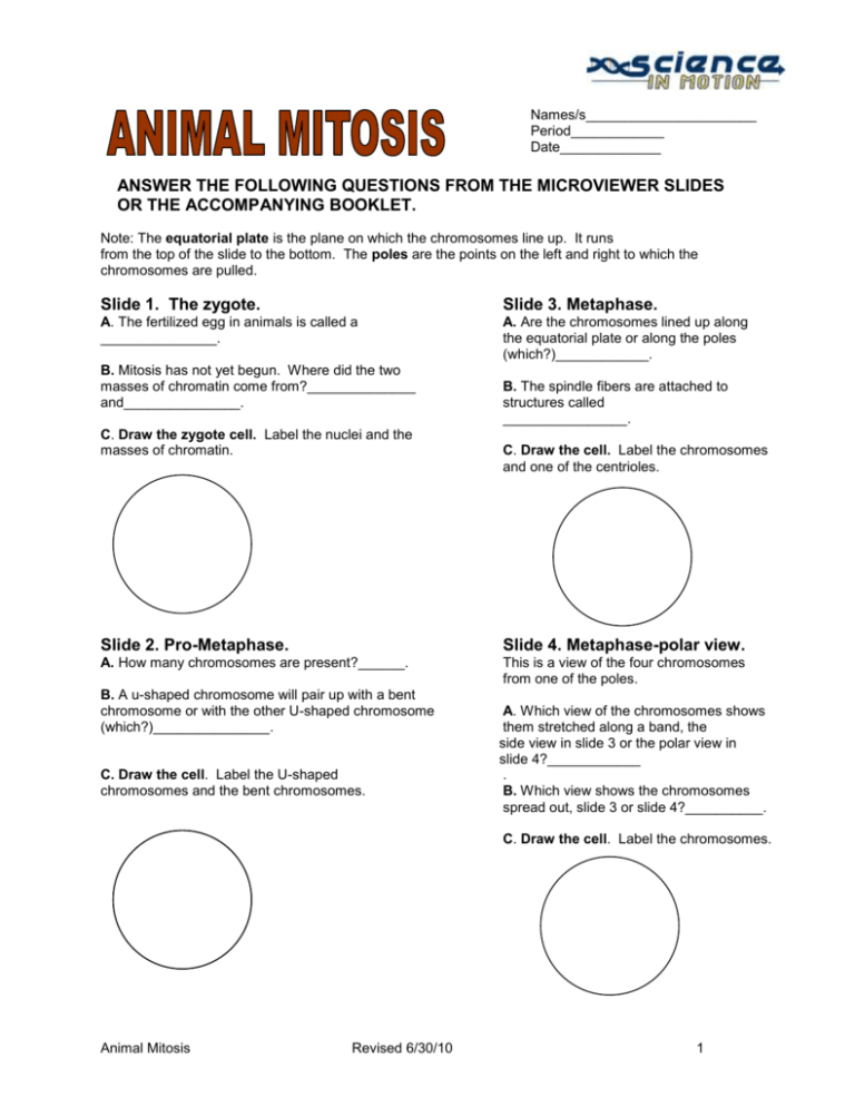
Names/s______________________ Period____________ Date_____________ ANSWER THE FOLLOWING QUESTIONS FROM THE MICROVIEWER SLIDES OR THE ACCOMPANYING BOOKLET. Note: The equatorial plate is the plane on which the chromosomes line up. It runs from the top of the slide to the bottom. The poles are the points on the left and right to which the chromosomes are pulled. Slide 1. The zygote. Slide 3. Metaphase. A. The fertilized egg in animals is called a _______________. A. Are the chromosomes lined up along the equatorial plate or along the poles (which?)____________. B. Mitosis has not yet begun. Where did the two masses of chromatin come from?______________ and_______________. C. Draw the zygote cell. Label the nuclei and the masses of chromatin. B. The spindle fibers are attached to structures called ________________. C. Draw the cell. Label the chromosomes and one of the centrioles. Slide 2. Pro-Metaphase. Slide 4. Metaphase-polar view. A. How many chromosomes are present?______. This is a view of the four chromosomes from one of the poles. B. A u-shaped chromosome will pair up with a bent chromosome or with the other U-shaped chromosome (which?)_______________. C. Draw the cell. Label the U-shaped chromosomes and the bent chromosomes. A. Which view of the chromosomes shows them stretched along a band, the side view in slide 3 or the polar view in slide 4?____________ . B. Which view shows the chromosomes spread out, slide 3 or slide 4?__________. C. Draw the cell. Label the chromosomes. Animal Mitosis Revised 6/30/10 1 Science in Motion Juniata College Slide 5. Early Anaphase. Slide 7. Telophase. Each of the four chromosomes duplicated much earlier, but the process was not visible, Now eight chromosomes are visible. The two sets of chromosomes are now completely separated. A. How many sets of chromosomes are beginning to form?__________. A. Earlier the chromosomes divided (duplicated). What is dividing now? ________________. B. How many chromosomes are in each set? _________. B. Draw the cell. Label the chromosomes, spindle fibers, and the centrioles. C. Draw the cell. Label the sets of chromosomes. Slide 6. Anaphase. Slide 8. Late Telophase. A. What structures are pulling the two sets of chromosomes apart?___________________. A. MItosis began with a single zygote cell. How many cells are present at late telophase?_______. B. What are the 'beaded' structures on the chromosomes?______________. C. Draw the cell. Label the chromosomes, and spindle fibers. B. The chromosomes are still fat and thick. What will happen to them after cell division is over?__________________________. C. Draw the cells. Label the chromosomes. These slides show the stages of mitosis in the roundworm, Ascaris. It is easier to follow mitosis in Ascaris than in humans, because the worm has only four diploid chromosomes and we have 46. All the views are at a magnification of 750x. Animal Mitosis Revised 6/30/10 2
