Suppl. Material
advertisement
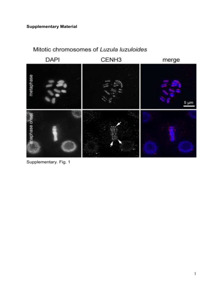
Supplementary Material Supplementary. Fig. 1 1 Supplementary Fig. 2 2 Supplementary Fig. 1 Morphology of mitotic L. luzuloides chromosomes during metaphase and at metaphase/anaphase transition. DAPI stained chromosomes are shown in blue and CENH3 signals are in red. Although the relatively small L. luzuloides chromosomes formed at metaphase/anaphase transition an indistinguishable mass of chromosomes, bending of chromosomes was indirectly visible via curved linear CENH3 signals (arrows) at this stage. Supplementary Fig. 2 Mitotic metaphase chromosomes of L. luzuloides after FISH (a, b) with 45S rDNA (green) and Arabidopsis-type telomere probes (red). DAPI stained chromosomes are shown in blue. (b) Notably, interstitial telomeric sites were detected on some chromosomes (marked by arrows) when CCD camera was long time exposed. Supplementary Video 1 Picture rotation of CENH3- and DAPI-labeled metaphase chromosomes of L. elegans recorded with deconvolution fluorescent light microscopy. Picture rotation visualizes a longitudinal groove along the poleward surface of each sister chromatid in DAPI (yellow). Linear-like CENH3 signal pairs (yellow) demonstrating localization on both sister chromatids colocalizing with the groove. Note that terminal chromosomal ends are groove- and CENH3-free. Supplementary Video 2 Picture rotation of CENH3-labeled chromosomes of L. elegans at metaphase/anaphase transition recorded with deconvolution fluorescent light microscopy. Curved linear CENH3 signals (yellow) indicating chromosome bending during metaphase/anaphase transition. 3
