Immunology - University of New Haven
advertisement
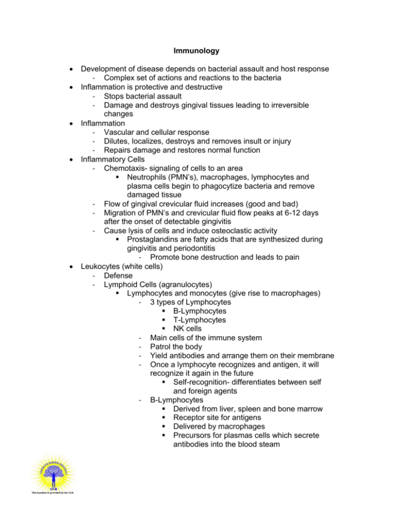
Immunology This handout is provided by the CLR. Development of disease depends on bacterial assault and host response - Complex set of actions and reactions to the bacteria Inflammation is protective and destructive - Stops bacterial assault - Damage and destroys gingival tissues leading to irreversible changes Inflammation - Vascular and cellular response - Dilutes, localizes, destroys and removes insult or injury - Repairs damage and restores normal function Inflammatory Cells - Chemotaxis- signaling of cells to an area Neutrophils (PMN’s), macrophages, lymphocytes and plasma cells begin to phagocytize bacteria and remove damaged tissue - Flow of gingival crevicular fluid increases (good and bad) - Migration of PMN’s and crevicular fluid flow peaks at 6-12 days after the onset of detectable gingivitis - Cause lysis of cells and induce osteoclastic activity Prostaglandins are fatty acids that are synthesized during gingivitis and periodontitis - Promote bone destruction and leads to pain Leukocytes (white cells) - Defense - Lymphoid Cells (agranulocytes) Lymphocytes and monocytes (give rise to macrophages) - 3 types of Lymphocytes B-Lymphocytes T-Lymphocytes NK cells - Main cells of the immune system - Patrol the body - Yield antibodies and arrange them on their membrane - Once a lymphocyte recognizes and antigen, it will recognize it again in the future Self-recognition- differentiates between self and foreign agents - B-Lymphocytes Derived from liver, spleen and bone marrow Receptor site for antigens Delivered by macrophages Precursors for plasmas cells which secrete antibodies into the blood steam - This handout is provided by the CLR. Stimulate other T-cells so immune system can grow - T-Lymphocytes Helper T cells - Assist B- cells in the production of antibodies - Stimulate B-cells to differentiate into plasmas cells which produce antibodies Cytotoxic T cells - Stimulate cytotoxic activity in cells such as macrophages - NK Cells Produce antibodies Stimulate microcidal effects of the immune response through the production of a variety of substances Effective against viruses and tumor cells Macrophages - Differentiate from monocytes - Phagocytic cell - “big eaters” - Attack antigen and present it to the lymphocyte - Antigen processed by macrophages » development of NK cells - In connective tissues that phagocytize bacteria but produce enzymes that degrade collagen B. Granulocytes Polymorphonuclear leukocytes (PMN’s) - Neutrophils - Major phagocytic cell - First line of defense Once of the first cells recruited to the site of inflammation - 70% of circulating leukocytes Mast Cells - Located in the tissue - Cytoplasm loaded with granules that contain histamine and heparin - Key players in the initiation of inflammation Basophils - Rarest leukocyte (less than 1%) - Small and rich in granules - Bi or tri lobed nucleus which is hard to see due to granules Eosinophils This handout is provided by the CLR. Attack parasites and phagocyte antigen-antibody complexes Effector Molecules - Antibodies (5 Classes) IgG- in blood and extravascular fluids, neutralize toxins and enhance phagocytosis, 80% of all antibody in serum IgM- develops early in immune system, early activation of complement IgE- involved in acute allergic reactions, reaction with antigen leads to histamine release IgD- low levels, important in triggering immune response IgA- in saliva, tears and bodily fluids, activates complement and interferes with bacterial adhesion, in gingival fluid (different than secretory IgA which does not play a role in perio disease and does not enter perio pockets) - Complement Made of proteins and glycoproteins Account for 10% of proteins in human serum Mediate degranulation of mast cells Generate chemotactic factors that attract PMN’s, macrophages and other leukocytes to inflamed areas » increases phagocytosis of bacteria Cascading reaction- once it begins it grows until completion - Allows for amplification of immune response Reaction can lead to tissue destruction due to release of toxins by bacteria, release of histamine and tissues that cause collagen destruction and elastin destruction - Opsonization- process of phagocytosis is more effective if opsonins are present Opsonins are antibodies or complements that bind to the surface of bacteria Coat the bacteria so they are easier identified by PMN’s and can be “swallowed” - Cytokines Chemical substances that provide communication between cells Produced by stimulated immune cells Assist in the development and regulation of activity of the immune effector cells Director in the complex communication system Contribute to periodontal bone resorption - Stimulate an increase in the number of osteoclasts - Stimulate cells to secrete prostaglandins Hypersensitivity Reactions - Overreactions by the immune system - This handout is provided by the CLR. Usually protective in nature but may cause periodontal tissue destruction - Type 1- Anaphylaxis Generalized or localized Seen in individuals with food or drug allergies - Type 2- Cytotoxic Results in breakdown of tissues or blood cells Not seen in perio or gingivitis In other oral diseases such as pemphigus - Type 3- Arthus Occurs when high level of antigen remain without being eliminated Occurs around small blood vessels Activates compliment and can cause extensive tissue damage - Type 4- Cell-Mediated or Delayed Seen in second exposures to an allergic agent T-lymphocytes after being sensitized to a specific antigen, undergo changes which increase the number of immunocompetent cells sensitive to the agent Calculus - Also called tartar - Supragingival most common on lower anterior lingual surfaces and maxillary first molar buccal surfaces Due to proximity to salivary glands - Contributes to perio disease but is not the causative agent Occurs when plaque is mineralized which occurs within 2448 hours - Matures in 12 days
![Immune Sys Quiz[1] - kyoussef-mci](http://s3.studylib.net/store/data/006621981_1-02033c62cab9330a6e1312a8f53a74c4-300x300.png)
