Comparing Cell Types OBJECTIVES After completing this lab you
advertisement
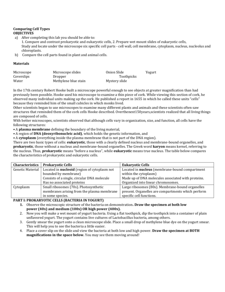
Comparing Cell Types OBJECTIVES a) After completing this lab you should be able to: 1. Compare and contrast prokaryotic and eukaryotic cells, 2. Prepare wet mount slides of eukaryotic cells, Study and locate under the microscope six specific cell parts - cell wall, cell membrane, cytoplasm, nucleus, nucleolus and chloroplasts. b) Compare the cell parts found in plant and animal cells. Materials Microscope Coverslips Water Microscope slides Dropper Methylene blue stain Onion Slide Toothpicks Mystery slide Yogurt In the 17th century Robert Hooke built a microscope powerful enough to see objects at greater magnification than had previously been possible. Hooke used his microscope to examine a thin piece of cork. While viewing this section of cork, he observed many individual units making up the cork. He published a report in 1655 in which he called these units “cells” because they reminded him of the small cubicles in which monks lived. Other scientists began to use microscopes to examine many different plants and animals and these scientists often saw structures that reminded them of the cork cells Hooke described. Overthenext150years,scientists realized that all living things are composed of cells. With better microscopes, scientists observed that although cells vary in organization, size, and function, all cells have the following structures: • A plasma membrane defining the boundary of the living material, • A region of DNA (deoxyribonucleic acid), which holds the genetic information, and • A cytoplasm (everything inside the plasma membrane that is not part of the DNA region). There are two basic types of cells: eukaryotic, those with a clearly defined nucleus and membrane-bound organelles, and prokaryotic, those without a nucleus and membrane-bound organelles. The Greek word karyon means kernel, referring to the nucleus. Thus, prokaryotic means “before a nucleus”, while eukaryotic means true nucleus. The table below compares the characteristics of prokaryotic and eukaryotic cells. Characteristics Genetic Material Prokaryotic Cells Eukaryotic Cells Located in nucleoid (region of cytoplasm not Located in nucleus (membrane-bound compartment bounded by membrane) within the cytoplasm) Consists of a single, circular DNA molecule Made up of DNA molecules associated with proteins. Has no associated proteins Organized into linear chromosomes. Cytoplasm Small ribosomes (70s). Photosynthetic Large ribosomes (80s). Membrane-bound organelles membranes arising from the plasma membrane present. Organelles are compartments which perform in some species. specific cell functions. PART I: PROKARYOTIC CELLS (BACTERIA IN YOGURT) 1. Observe the microscopic structure of the bacteria on demonstration. Draw the specimen at both low power (40x) and medium (100x) OR high power (400x). 2. Now you will make a wet mount of yogurt bacteria. Using a flat toothpick, dip the toothpick into a container of plain unflavored yogurt. The yogurt contains live cultures of Lactobacillus bacteria, among others. 3. Gently smear the yogurt onto a clean microscope slide. Place a small drop of methylene blue dye on the yogurt smear. This will help you to see the bacteria a little easier. 4. Place a cover slip on the slide and view the bacteria at both low and high power. Draw the specimen at BOTH magnifications in the space below. You may see them moving around! 5. Clean and dry your slide and return it to the supply area when you are finished. Examine the drawing of the bacterium Escherichia coli below. The cell has a cell wall, a structure different from the wall of plant cells but serving the same primary function. The plasma membrane is flat against the cell wall and may be difficult to see. Look for two components in the cytoplasm: the small block dots called ribosomes give the cytoplasm its granular appearance; the nucleoid, a relatively electron-transparent region (appears light) containing fine threads of DNA. Label the structures highlighted structures discussed above on the diagram below. Part II: Eukaryotic Cells – Animal Cells (Cheek Cells) 1. Obtain a clean toothpick, slide, and coverslip. 2. Use a clean toothpick to gently scrape the inside of your cheek. (You will not be able to see anything on the toothpick) 3. Add ONE drop of methylene blue from the pipet to the slide. Roll the toothpick with your cheek cells in the drop. Add a cover slip and throw the toothpick in the trash. 4. Locate the cheek cells using low power, then switch to middle power, then switch to high power. Find the nucleus, a round centrally located body within each cell. Draw the specimen at both low power (40x) and medium (100x)/high power (400x) in the space below. 6. Carefully draw several cells as they appear under the microscope. Label the cytoplasm, nucleus, and plasma membrane. Record the magnification used. Locate and examine cells that are separated from one another rather than those that are in clumps. Part II: (A) Eukaryotic Cells – Plant Cells (ONION CELLS) 1. An onion epidermal cell slide has been prepared. 2. Observe the wet mount with your microscope under low power, then switch to middle power, then switch to high power. 3. The nucleus will be a large sphere within the cytoplasm. Examine the nucleus carefully and you will spot several nucleoli inside the nucleus. Nucleoli are the areas within the nucleus were RNA (ribonucleic acid) is produced. The rest of the nucleus is largely DNA. 4. Look for oil droplets in the form of granular material within the cytoplasm. The droplets are a form of stored food for the cell. 5. Carefully draw several onion epidermal cells. Draw the specimen at both low power (40x) and medium (100x)/high power (400x) in the space below. Label the oil droplets, nucleus, cell wall, cell membrane, and cytoplasm. Part II: (B) Eukaryotic Cells--Plant Cells (LETTUCE CELLS) 6. Now prepare a slide of green leaf lettuce. You will need to tear off a small piece of the leaf. 8. Place a small drop of water on the center of a slide. Put the leaf piece in the drop of water. 7. Place a cover slip on the specimen. Look carefully for chloroplasts and the central vacuole in the lettuce leaf cells. 8. Carefully draw several lettuce leaf cells. Draw the specimen at both low power (40x) and medium (100x)/high power (400x) in the space below). Label the chloroplasts, central vacuole, nucleus (if seen; it may be hard to see), cell wall, cell membrane, and cytoplasm. 9. Clean off your slides, dry them and return them to the supply area. Analysis Part A ONION CELLS-Cell wall, Cell membrane, Nucleus, Cytoplasm 1.Describe the shape of an onion cell: 2. a) Are onion cells produced by plants or animals? b) How do you know? 3.What is the function of a cell’s nucleus? 4.Why were the cells stained? CHEEK CELLS-The Cell Membrane, Cytoplasm, and Nucleus 1. Describe the shape of a cheek cell: 2. a) Are cheek cells produced by plants or animals? b) How do you know? 3. Are cheek cells alive? LETTUCE CELLS-Cell Wall, Cell Membrane, Cytoplasm, and Chloroplast 1.Describe the shape of a lettuce cell: 2. a) Does lettuce have plant or animal cells? b) Provide two pieces of evidence to defend your answer in 2a. 3.Describe the a) color of the chloroplast 4.What is the function of the chloroplast? Analysis Part B 1) Determine if each of the following characteristics is true of Prokaryotic cells, Eukaryotic cells, or Both cell types. ______ No membrane-bound nucleus ______ Membrane-bound nucleus ______ No membrane-bound organelles ______ Contains membrane-bound organelles ______ Small ribosomes present ______ Large ribosomes present ______ Cell membrane present ______ Chromosomes present ______ Cytoplasm present ______ Mitochondria, endoplasmic reticulum, Golgi, and vacuoles present ______ Genetic material is linearly arranged 2) How does the size of the bacterial cell observed compare with the size of the eukaryote cells you viewed? 3) Name 4 structures common to all cells. ________________________________________ _________________________________________ ________________________________________ _________________________________________ 4)Name 3 differences between plant and animal cells. ________________________________________________________________________________________ ________________________________________________________________________________________ 5) Mitochondria and chloroplasts have their own DNA and ribosomes. What does this tell you about their possible origin? ________________________________________________________________________________________ _______________________________________________________________________________________


