SUPPLEMENTARY INFORMATION
advertisement
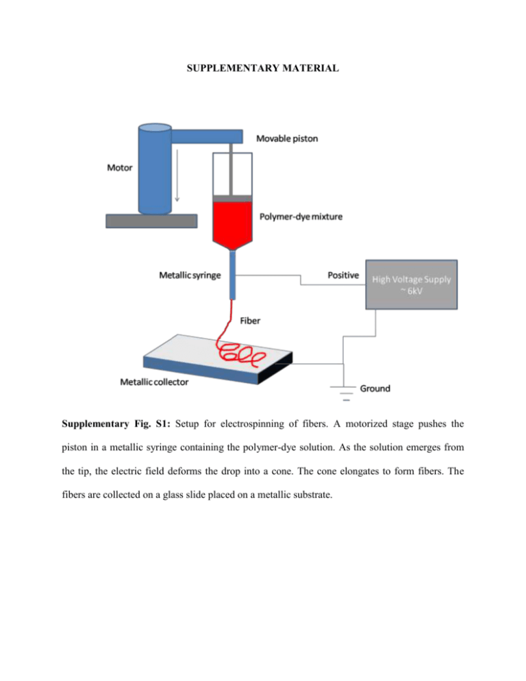
SUPPLEMENTARY MATERIAL Supplementary Fig. S1: Setup for electrospinning of fibers. A motorized stage pushes the piston in a metallic syringe containing the polymer-dye solution. As the solution emerges from the tip, the electric field deforms the drop into a cone. The cone elongates to form fibers. The fibers are collected on a glass slide placed on a metallic substrate. Supplementary Fig. S2: Confocal image of a typical fiber with the fluorescence spectrum from the spot labeled as 1. Supplementary Fig. S3: Intensity of emission for two different pump powers-one below threshold (red curve) and above threshold (blue curve). The emission intensity increases rapidly at threshold and a simultaneous narrowing of the spectrum can be observed. Conducting needle Field lines Supplementary Fig. S4: Field lines from the conducting needle towards the collecting substrate. References: 1. Li et al, Nano Letters 3, 1167-1171, 2003.


