REV metal nanohelices room-temp growth-supp
advertisement
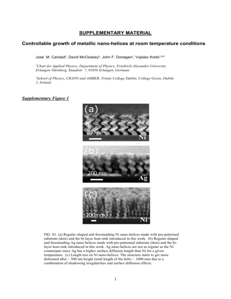
SUPPLEMENTARY MATERIAL Controllable growth of metallic nano-helices at room temperature conditions José M. Caridad2, David McCloskey2, John F. Donegan2, Vojislav Krstić1,2* 1 Chair for Applied Physics, Department of Physics, Friedrich-Alexander-University Erlangen-Nürnberg, Staudtstr. 7, 91058 Erlangen, Germany 2 School of Physics, CRANN and AMBER, Trinity College Dublin, College Green, Dublin 2, Ireland Supplementary Figure 1 FIG. S1. (a) Regular-shaped and freestanding Ni nano-helices made with pre-patterned substrate (dots) and the bi-layer heat-sink introduced in this work. (b) Regular-shaped and freestanding Ag nano-helices made with pre-patterned substrate (dots) and the bilayer heat-sink introduced in this work. Ag nano-helices are not as regular as the Ni counterpart since Ag has a higher surface diffusion length than Ni for a given temperature. (c) Length test on Ni nano-helices. The structure starts to get more deformed after ~ 500 nm height (total length of the helix ~ 1000 nm) due to a combination of shadowing irregularities and surface diffusion effects. 1 Supplementary Figure 2 FIG. S2. SEM image of Ni nano-helices under non-optimized growth on a transparent substrate composed of SiO2 and a thin film (150 nm) of ITO as a heat-sink layer. Note that this image was not taken at a normal angle with respect to the helical axis. Supplementary Table S1 Sample (Metal) pitch, 𝑝 (nm) diameter, 𝐷 (nm) nano-helix height, ℎ (nm) no. of turns, 𝑁 S1(Ni) 325 ± 9 169 ± 9 400 ± 20 1.23 S2 (Ni) S3(Ni) S4 (Ni) S5 (Ni) S6 (Ag) S7 (Ag) S8 (Ag) S9 (Ag) 130 ± 6 128 ± 9 63 ± 6 128 ±10 130 ± 9 140 ± 11 127 ± 9 127 ± 10 102 ± 6 72 ± 9 59 ± 6 72 ± 7 75 ± 7 103 ± 9 72 ± 7 72 ± 6 242 ± 15 390 ± 16 234 ± 20 275 ± 15 300 ± 16 300 ± 13 268 ± 12 238 ± 11 1.86 3.04 3.70 2.3 2.3 2.1 2.11 1.88 Table S1 Structural helix parameters of the grown structures, showing a variation of pitch and diameter below 10%. 2 Optical activity measurements The optical activity of the metallic nano-helices was acquired in transmission using a commercial spectrometer with liquid nitrogen cooled CCD detector (Horiba Triax 180) to record the spectral response of the sample in the range from 400 to 1000 nm. A Xe discharge lamp was used as a light source and a linear polarizer and a Fresnel romb were inserted to produce circular right- or lefthanded polarized light, RHC and LHC respectively. The white light was focused on the sample through a 20x NA=0.4 microscope objective and the areas of interest of the sample are selected with a 2 axis variable slit installed in the set-up. 3


