P wave corresponds to the atria
advertisement
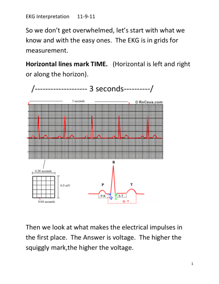
EKG Interpretation 11-9-11 So we don’t get overwhelmed, let’s start with what we know and with the easy ones. The EKG is in grids for measurement. Horizontal lines mark TIME. (Horizontal is left and right or along the horizon). /-------------------- 3 seconds----------/ Then we look at what makes the electrical impulses in the first place. The Answer is voltage. The higher the squiggly mark,the higher the voltage. 1 EKG Interpretation 11-9-11 The vertical lines mark VOLTAGE. Since we know that during several of these grids (that mark time) we should be seeing several P QRS and T complexes showing the voltage. However, if we see an EKG strip that looks like this: There is no voltage. There is no heartbeat over a significant period of time. This cardiac standstill. This is asystole! YIKES! A normal EKG has all of the waves we learned about and it looks like this: 2 EKG Interpretation 11-9-11 The first little bump is the P Wave and looks rounded, like it should. It is followed by the big upside down QRS complex and it looks fine. Then the last bump is the T wave and it is fine. We don’t really see the U wave here and since we aren’t telemetry techs, we aren’t going to worry too much about the U wave. So the strip above and below this text represents normal sinus rhythm. (Isn’t it pretty?!) normalsinus.blogspot.com/ What about identifying fast and slow heart beats? Just don’t panic when you look at an ekg strip and jump to conclusions! Remember to look at how even the QRS complexes are to one another (REGULAR) and how many of them there are. 3 EKG Interpretation 11-9-11 On this strip we see that the P, QRS and T waves all look pretty normal. In addition, the distance the QRS complexes are apart, is pretty even or regular. So, the main problem with this Ekg strip is that the heart is beating too slow. So we know that this is bradycardia. A person may panic when looking at this strip and say that the T waves and P waves are close together or whatever. As I said before, just don’t panic. Count the QRS complexes. Gee, there sure are a LOT of them! This person is tachycardic. Ask yourself, what kind of reasons could the heart have for suddenly going fast. We know that if the cells of the body do not get enough oxygen, they put out a signal. If, for example, there has been a big loss of blood, the body 4 EKG Interpretation 11-9-11 will not have enough red blood cells to carry oxygen. The heart responds by circulating around what rbc’s are left, in a big dang hurry! Yes, hemorrhage is one of the reasons a heart may speed along. Great, now we know normal sinus rhythm, asytole, bradycardia and tachycardia. Next, we will show the wild roller coaster which is Ventricular Tachycardia because V tach is easy to recognize. It looks like this: and this: and this: Ventricular Tachycardia 5 EKG Interpretation 11-9-11 Why is it far worse for the Ventricles to quiver and be ineffective than if the Atria quiver and are ineffective? I mean, after all, a person with A fib can walk in a clinic, sign a consent form, get medicated, and get shocked back to a normal rhythm, but a guy with V Fib isn’t going to be talking to anyone or awake enough to be signing any consent forms! The answer to this question is that the atria are small and do not contribute as much to pumping the heart as the ventricles do! The only good thing you can say about V tach is that at least there IS some cardiac rhythm in order to shock it into a normal sinus rhythm. With asytole,(cardiac standstill), the experts say that defibrillation does not work and that when asytole occurs outside of the hospital, only about 2% of its victims survive! ============================================ Now let’s move on to P Waves: 6 EKG Interpretation 11-9-11 The P wave corresponds to the atria. An absent P Wave means something is wrong with the atria such as atrial fibrillation. Absent P Wave looks like this: Absent. Absent P waves have four causes: Atrial fibrillation Sinus arrest Sinoatrial block Hyperkalaemia What if there are TWO P Waves? Two P waves mean 3rd degree heart block which must be treated immediately. A pacemaker or defibrillator may be necessary. Two P Waves. Two P Waves = 3rd Degree heart block. 7 EKG Interpretation 11-9-11 www.emsvillage.com/articles/article.cfm?id=684 3rd degree heart block 3rd degree heart block www.sciencephoto.com/image/272870/large/M4600... 8 EKG Interpretation 11-9-11 Why do they talk so much about measuring the PR interval? What is the PR interval anyway? The PR interval is the delay time. This is the time between the P Wave and the QRS complex. This is the time it takes the impulse to come from the atria and travel through the AV node and into the left bundle branches. This PR interval is a very short “time out”. Think of it like this: When it comes to the four chambers of the heart, the atria are the little guys. They are smaller than the ventricles. We will call them the Atria Brothers: Lefty and Righty. Being a good buddy to his small friends, “AV node” shows his concern for them by slowing down this lightening fast electrical impulse in order to give his friends a change to finish their job of pumping out their blood out of the top chambers of the heart and into the bottom chambers of the heart BEFORE the big strong Ventricle Brothers start in. The delay helps prevent kick back of blood. In fact, if it wasn’t for good old buddy “AV”, then the little Atria Brothers would get overwhelmed by the Ventricle Brothers. 9 EKG Interpretation 11-9-11 Or, to think of it another way, let’s say that “AV node” is a telegraph station. He gets the message from Master Node (SA node) that he is supposed to pass on, telling the big stagecoach to leave the station. But he knows that it would be better if his two small friends on horseback got to pull into the station and tie up their horses BEFORE the big coach takes off or else his little friends might get bucked off their horses. So, being the good buddy that he is, “AV” waits a fraction of a second before he passes the message on. This delay is only .12 to .20 of a second. (Like I said, it is REALLY fast!) However, if this delay is LONGER than .20 of a second, then the good old AV didn’t merely delay the message, he blocked it! If the delay is SHORTER than .12 seconds, the electrical impulse is not going through healthy and faithful friend “AV”. We know that if it goes too fast, then the electrical impulse is traveling from the SA node through another route and bypassing “AV”. In this strange route, the 10 EKG Interpretation 11-9-11 node there does NOT delay the message, which is going to be really hard on the Atria Brothers. Atrial Flutter Atrial flutter is an abnormality of the heart rhythm, resulting in a rapid and sometimes irregular heartbeat http://www.google.com/imgres?imgurl=http://www.learnekgs.com/AFlutter%2520LARGE.jpg&imgrefurl=http://www.12leads.com/atrialflutter.htm&usg=__JKMSzjz_AnPzCoV53fNgkjh0f0=&h=413&w=1778&sz=315&hl=en&start=6&zoom=1&tbnid=ulKcD1L0rqXbDM:&tbnh=35&tbnw=150&ei=_Ym6T t3RG7Pg2wX5mISvBw&prev=/search%3Fq%3Datrial%2Bflutter%26hl%3Den%26sa%3DX%26gbv%3D2%26tbm%3Disch%26prmd%3Divns&itbs= 1 Atrial flutter abnormal electrical path through the atria. This is why sometimes the solution is to cut the abnormal pathway (known as ablation). A. Fib www.pedcard.rush.edu/.../irregular-fastlrate.htm 11 EKG Interpretation 11-9-11 Atrial fibrillation is an irregular and often rapid heart rate that commonly causes poor blood flow to the body. The atria beat chaotic and irregular. Question: What causes wandering on a baseline ekg? Answer: sweat and lotion Question: What happens to the waveform on the ECG if a lead comes off the patient’s chest? 12 EKG Interpretation 11-9-11 Answer: This stops the monitoring process. The waveform will not be seen on the monitor. 13
