Cells: The Living Units Chapter 3 I.
advertisement
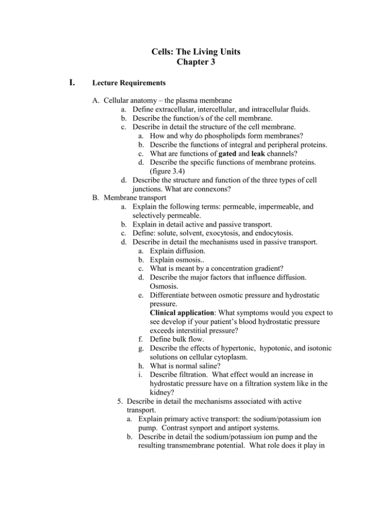
Cells: The Living Units Chapter 3 I. Lecture Requirements A. Cellular anatomy – the plasma membrane a. Define extracellular, intercellular, and intracellular fluids. b. Describe the function/s of the cell membrane. c. Describe in detail the structure of the cell membrane. a. How and why do phospholipds form membranes? b. Describe the functions of integral and peripheral proteins. c. What are functions of gated and leak channels? d. Describe the specific functions of membrane proteins. (figure 3.4) d. Describe the structure and function of the three types of cell junctions. What are connexons? B. Membrane transport a. Explain the following terms: permeable, impermeable, and selectively permeable. b. Explain in detail active and passive transport. c. Define: solute, solvent, exocytosis, and endocytosis. d. Describe in detail the mechanisms used in passive transport. a. Explain diffusion. b. Explain osmosis.. c. What is meant by a concentration gradient? d. Describe the major factors that influence diffusion. Osmosis. e. Differentiate between osmotic pressure and hydrostatic pressure. Clinical application: What symptoms would you expect to see develop if your patient’s blood hydrostatic pressure exceeds interstitial pressure? f. Define bulk flow. g. Describe the effects of hypertonic, hypotonic, and isotonic solutions on cellular cytoplasm. h. What is normal saline? i. Describe filtration. What effect would an increase in hydrostatic pressure have on a filtration system like in the kidney? 5. Describe in detail the mechanisms associated with active transport. a. Explain primary active transport: the sodium/potassium ion pump. Contrast synport and antiport systems. b. Describe in detail the sodium/potassium ion pump and the resulting transmembrane potential. What role does it play in muscle contraction and the transmission of information along nerve tissue? c. Vesicular transport 1) Contrast exocytosis with endocytosis. 2) Explain the relationship between phagosomes, phagolysosomes, and phagocytosis. 3) Describe pinocytosis. 4) Explain receptor-mediated endocytosis. C. Cellular structures a. The cytoplasm a. Differentiate between cytosol and organelles b. Which organelles are membranous? Which are nonmembranous? c. Describe the cytoskeleton. d. Describe the structure and function of all cellular organelles. b. The nucleus a. Describe the structure of the chromosome. Relate the nucleosome, centromere, and chromatin. b. Describe the structure of DNA. c. What is the “genetic code?” d. Explain DNA replication. e. Describe the structure of the nucleolus. What is its function? Does it have DNA or RNA in its structure? What type cells normally have a greater concentration of nucleoli? D. Protein synthesis a. Explain protein synthesis. b. Describe how transcription and translation function in protein synthesis. c. Explain triplets, codons, and anticodons d. Explain the roles of mRNA and tRNA in protein synthesis. e. What role does ribosomal RNA play in protein synthesis? E. Cell growth and reproduction 1. Describe the cell cycle. a. Describe the sub-phases of interphase . What significant event takes place during S-subphase? Describe this process. b. Describe the events that are associated with each of the four phases of mitosis. 2. Explain cytokinesis *******In lab you will have to be able to identify each phase******** F. Problems 1. Replicate this DNA strand. ATC GAT ----------AGG HHH HHH HHH TAG CTA ----------TCC 2. Transcribe the following gene. AAC GAT --------AAT TCG 3. Translate the following mRNA. AUG UUU ------- UAA GUU II. Laboratory exercise A. Make a slide of a human cheek cell and observe its cellular components. B. Observe the slides associated with the cell cycle.
