CHRONIC DIARRHEA: Beyond Bugs and Drugs OBJECTIVES
advertisement
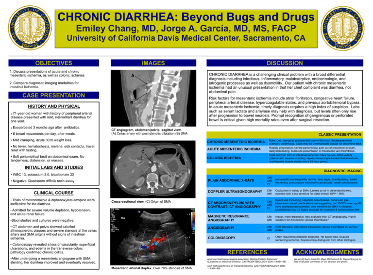
CHRONIC DIARRHEA: Beyond Bugs and Drugs Emiley Chang, MD, Jorge A. García, MD, MS, FACP University of California Davis Medical Center, Sacramento, CA OBJECTIVES 1. Discuss presentations of acute and chronic mesenteric ischemia, as well as colonic ischemia. IMAGES CHRONIC DIARRHEA is a challenging clinical problem with a broad differential diagnosis including infectious, inflammatory, malabsorptive, endocrinologic, and iatrogenic processes as well as dysmotility. Our patient with chronic mesenteric ischemia had an unusual presentation in that her chief complaint was diarrhea, not abdominal pain. A 2. Compare diagnostic imaging modalities for intestinal ischemia. CASE PRESENTATION HISTORY AND PHYSICAL Risk factors for mesenteric ischemia include atrial fibrillation, congestive heart failure, peripheral arterial disease, hypercoagulable states, and previous aortobifemoral bypass. In acute mesenteric ischemia, timely diagnosis requires a high index of suspicion. Labs such as serum lactate and amylase may help with diagnosis, but levels often only rise after progression to bowel necrosis. Prompt recognition of gangrenous or perforated bowel is critical given high mortality rates even after surgical resection. B 71-year-old woman with history of peripheral arterial disease presented with mild, intermittent diarrhea for one year. Exacerbated 3 months ago after antibiotics. • 6 bowel movements per day, after meals. CT angiogram, abdomen/pelvis; sagittal view. (A) Celiac artery with post-stenotic dilatation (B) SMA • Mild cramping, acute 30 lb weight loss. • No fever, hematochezia, melena, sick contacts, travel, relief with fasting. DISCUSSION CLASSIC PRESENTATION Rare. Dull, cramping, postprandial pain in first hour. Subsequent food aversion, weight loss. Exam may be unremarkable except for abdominal bruit. Rapidly progressive, severe periumbilical pain out-of-proportion to exam, delayed bleeding. Subacute presentation in mesenteric vein thrombosis. Most common form of intestinal ischemia; many causes. Often elderly patients with nausea, vomiting, rapidly worsening left-sided abdominal pain, and frequent bloody stools over a 24-hour period. CHRONIC MESENTERIC ISCHEMIA C • Soft periumbilical bruit on abdominal exam. No tenderness, distension, or masses. ACUTE MESENTERIC ISCHEMIA COLONIC ISCHEMIA INITIAL LABS AND STUDIES DIAGNOSTIC IMAGING • WBC 13, potassium 3.0, bicarbonate 30 • Negative Clostridium difficile toxin assay CLINICAL COURSE • Trials of metronidazole & diphenoxylate-atropine were ineffective for the diarrhea. Cross-sectional view. (C) Origin of SMA • Admitted for severe volume depletion, hypotension, and acute renal failure. •Stool studies and cultures were negative. • CT abdomen and pelvis showed calcified atherosclerotic plaques and severe stenosis at the celiac artery and SMA origins without signs of intestinal ischemia. PLAIN ABDOMINAL X-RAYS CMI AMI CI Nonspecific and frequently normal. Ileus signs, thumbprinting, bowel thickening, pneumatosis intestinalis (advanced). Vessel calcifications. DOPPLER ULTRASONOGRAPHY CMI AMI Occlusions in celiac or SMA. Limited by air in distended bowels, operator skill. Less sensitive for distal emboli. NPV ~ 99%. CT ABDOMEN/PELVIS WITH CONTRAST; CT ANGIOGRAPHY CMI AMI CI Bowel wall thickening, intestinal pneumatosis, portal vein gas. Mesenteric vessel calcifications are suggestive, but 10-20% over age 65 have asymptomatic stenosis. Very sensitive for SMV thrombosis. Multidetector row CT highly accurate for occlusive & nonocclusive AMI. MAGNETIC RESONANCE ANGIOGRAPHY CMI AMI Newer, more expensive, less available than CT angiography. Highly sensitive for mesenteric venous thrombosis? ANGIOGRAPHY CMI AMI Gold standard. Can detect mesenteric venous thrombosis on venous phase. CI Often required to establish diagnosis. No bowel prep, to avoid worsening ischemia. Biopsies help distinguish from other etiologies. COLONOSCOPY • Colonoscopy revealed a loss of vascularity, superficial ulcerations, and edema in the transverse colon; pathology confirmed chronic colitis. •After undergoing a mesenteric angiogram with SMA stenting, her diarrhea improved and eventually resolved. Mesenteric arterial duplex. Over 70% stenosis of SMA. REFERENCES ACKNOWLEDGMENTS American Gastroenterological Association Medical Position Statement: Guidelines on Intestinal Ischemia. GASTROENTEROLOGY 2000;118:951–953 We would like to thank Dr. Maya Mitchell and Dr. Ripple Sharma for their invaluable comments on our abstract and poster. AGA Technical Review on Intestinal Ischemia. GASTROENTEROLOGY 2000; 118:954–968

![Paper_Prof_Wang_final1[1]](http://s3.studylib.net/store/data/005836194_1-85fb8d8882c087decd1a6d9c9fdc99c0-300x300.png)
