Embryology
advertisement
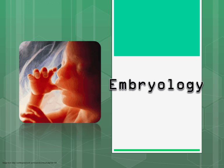
Image from: http://earthhopenetwork.net/forum/showthread.php?tid=706 = The branch of biology that studies the formation and early development of living organisms Embryo (implantation to 8 weeks) Fetus (after 8 weeks) Image from: http://www.amazon.com/Nova-Miracle-Life-Lennart-Nilsson/dp/6302895189; A. Fertilization B. Implantation C. Gastrulation D. Cell differentiation Images from: http://en.wikipedia.org/wiki/Fertilisation; http://www.lifenews.com/2011/12/05/pregnancy-begins-at-conceptionfertilization-not-implantation/ http://www.bio.miami.edu/dana/106/106F05_4.html; ; 1. =Egg and sperm come together to form a fertilized egg (called a … ) Fertilization Human Zygote Click for Video Images from: http://en.wikipedia.org/wiki/Fertilisation; http://php.med.unsw.edu.au/embryology/index.php?title=Fertilization; http://www.exploratorium.edu/imaging_station/gallery.php?Asset=Zebrafish%20development&Group=&Category=Zebrafish&Section=Introduction 2. After fert., before implantation . . . a. The zygote begins to divide i. First few mitotic cell divisions are called cleavage 2. After fert., before implantation . . . a. The zygote begins to divide i. First few mitotic cell divisions are called cleavage b. Soon a solid ball of cells is formed, called a .... A scanning electron micrograph of a human embryo at the eight-cell stage (day three). Image from: http://www.biologyreference.com/Co-Dn/Development.html 2. After fert., before implantation . . . a. The zygote begins to divide i. First few mitotic cell divisions are called cleavage b. Soon a solid ball of cells is formed, called a .... morula c. Next, the morula will change into a fluid filled ball of cells, called a. . . Click on image. Video at bottom left of page. Image and video from: http://worms.zoology.wisc.edu/dd2/echino/cleavage/blastula/blastula.html 1. The blastula will attach to the uterine wall. We call this attachment IMPLANATION Image from: http://www.implantationbleedinginsight.com/ Implantation Embryo 1. The process in which the cells of the blastocyst develop into three germ layers to form a . . . . Images from: http://biologieprojekt2009.wordpress.com/von-der-zygote-zum-mehrzeller/; Images from: http://biologieprojekt2009.wordpress.com/von-der-zygote-zum-mehrzeller/; http://moodle.urbandale.k12.ia.us/mod/slideshow/view.php?id=1932&img_num=2 2. The formation of 3 germ layers a. Endoderm • Inner-most layer • (digestive tract) b. Mesoderm • Middle layer • (internal organs) c. Ectoderm • Outer-most layer • (skin and nervous system) Images from: http://www.childrenscolorado.org/wellness/info/kids/21825.aspx; https://marketplace.secondlife.com/p/Beating-Anatomical-Heart-Animation-Sound/833619?id=833619&slug=Beating-Anatomical-Heart-Animation-Sound; http://www.rocketgirls.org/8-steps-for-beautifulglowing-skin-cleanse.html; http://biologieprojekt2009.wordpress.com/von-der-zygote-zum-mehrzeller/; Zygote Morula Blastula Gastrula 1. Once 3 germ cell layers formed in gastrula, then . . . Cell differentiation (or giving cells specific jobs) begins . . . a. 1st = neurulation i. Development of nervous system ii. Occurs soon after gastrulation is complete b. Organogenesis = organs start to form c. Morphogenesis = limbs start to assume shape A. Membranes form to protect and nourish the developing embryo. 1 = amnion membrane forms the amniotic sac (fluid filled cushions embryo) Image from: http://www.billcasselman.com/dictionary_of_medical_derivations/dmd_eight.htm 2 = chorion forms outer most membrane. This combined with uterine lining form the organ called the placenta Image from: http://bohone.wikispaces.com/Group10 The PLACENTA is a maternal-fetal organ which begins developing at implantation of the blastocyst Image from: http://embryology.med.unsw.edu.au/Medicine/BGDlabplacenta.htm = unspecialized cells that have the potential to differentiate 1. Totipotent Stem Cells These are the most versatile of the stem cell types. When a sperm cell and an egg cell unite, they form a one-celled fertilized egg. This cell is totipotent, meaning it has the potential to give rise to any and all human cells, such as brain, liver, blood or heart cells. It can even give rise to an entire functional organism. The first few cell divisions in embryonic development produce more totipotent cells. After four days of embryonic cell division, the cells begin to specialize into pluripotent stem cells. 2. Pluripotent Stem Cells These cells are like totipotent stem cells in that they can give rise to all tissue types. Unlike totipotent stem cells, however, they cannot give rise to an entire organism. On the fourth day of development, the embryo forms into two layers, an an outer layer which will become the placenta, and an inner mass which will form the tissues of the developing human body. These inner cells, though they can form nearly any human tissue, cannot do so without the outer layer; so are not totipotent, but pluripotent. As these pluripotent stem cells continue to divide, they begin to specialize further. Source: Brown University, Division of Biology and Medicine—Retrieved 6.7.11 from: http://biomed.brown.edu/Courses/BI108/BI108_2002_Groups/pancstems/stemcell/stemcellsclassversatility.htm 3. Multipotent Stem Cells These are less plastic and more differentiated stem cells. They give rise to a limited range of cells within a tissue type. The offspring of the pluripotent cells become the progenitors of such cell lines as blood cells, skin cells and nerve cells. At this stage, they are multipotent. They can become one of several types of cells within a given organ. For example, multipotent blood stem cells can develop into red blood cells, white blood cells or platelets 4. Adult Stem Cells An adult stem cell is a multipotent stem cell in adult humans that is used to replace cells that have died or lost function. It is an undifferentiated cell present in differentiated tissue. It renews itself and can specialize to yield all cell types present in the tissue from which it originated. So far, adult stem cells have been identified for many different tissue types such as hematopoetic (blood), neural, endothelial, muscle, mesenchymal, gastrointestinal, and epidermal cells. Source: Brown University, Division of Biology and Medicine—Retrieved 6.7.11 from: http://biomed.brown.edu/Courses/BI108/BI108_2002_Groups/pancstems/stemcell/stemcellsclassversatility.htm
