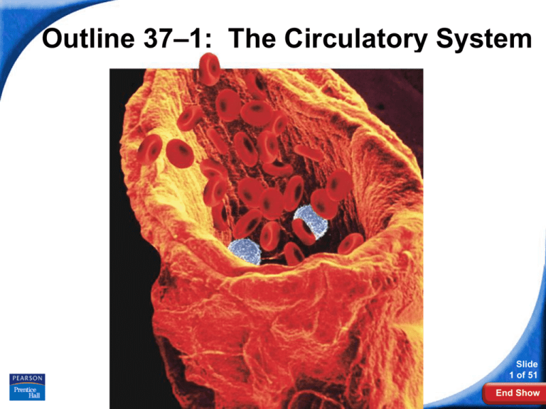
Outline 37–1: The Circulatory System
Slide
1 of 51
Copyright Pearson Prentice Hall
End Show
37–1 The Circulatory System
37–1 The Circulatory System
The circulatory system and respiratory system work
together to supply cells with the nutrients and oxygen
they need to stay alive.
a) The respiratory system:
● picks up the oxygen and absorbs it into
the blood.
● It changes oxygen-poor blood
(deoxygenated) into oxygen-rich
blood (oxygenated)
b) The circulatory system:
● then pumps the blood to the lungs &
rest of body
Slide
2 of 51
Copyright Pearson Prentice Hall
End Show
37–1 The Circulatory System
Functions of the Circulatory
System
Functions of the Circulatory System
Organisms with many cells need a way
to get oxygen & nutrients to each and
every cell of their body. The circulatory
system is the transport system of the
body that can do this.
Humans and other vertebrates have a
closed circulatory system, meaning
that the blood is always contained
within a system of vessels.
Copyright Pearson Prentice Hall
Slide
3 of 51
End Show
37–1 The Circulatory System
Functions of the Circulatory
System
The human circulatory system
consists of:
• the heart
• blood vessels
• blood
Slide
4 of 51
Copyright Pearson Prentice Hall
End Show
37–1 The Circulatory System
Blood Vessels
As blood flows through the
circulatory system, it moves
through three types of blood
vessels:
• arteries
• capillaries
• veins
Slide
5 of 51
Copyright Pearson Prentice Hall
End Show
37–1 The Circulatory System
Blood Vessels
Arteries
Large vessels that carry blood AWAY from
the heart to the tissues of the body are
called arteries.
Except for the pulmonary arteries, all
arteries carry oxygenated blood.
Arteries have thick muscular walls.
They contain the following tissues from
outside to inside: connective tissue, smooth
muscle, and endothelium.
Slide
6 of 51
Copyright Pearson Prentice Hall
End Show
37–1 The Circulatory System
Blood Vessels
Slide
7 of 51
Copyright Pearson Prentice Hall
End Show
37–1 The Circulatory System
Blood Vessels
Capillaries
The smallest of the blood vessels are
the capillaries. No cells are far from a
capillary.
Their walls are only one cell thick, and
most are so narrow that only one red
blood cell can pass through at a time.
Slide
8 of 51
Copyright Pearson Prentice Hall
End Show
37–1 The Circulatory System
Blood Vessels
The capillaries are exchange vessels:
They bring nutrients and oxygen to
the tissues of body
They absorb carbon dioxide and other
waste products from body cells and
bring these compounds away from cells
so the body can dispose of them.
Slide
9 of 51
Copyright Pearson Prentice Hall
End Show
37–1 The Circulatory System
Blood Vessels
Slide
10 of 51
Copyright Pearson Prentice Hall
End Show
37–1 The Circulatory System
Blood Vessels
Veins
Blood vessels that carry blood back to
the heart are called veins.
Except for the pulmonary veins, all
veins carry deoxygenated blood.
Veins have thinner walls than arteries,
containing less muscle than arteries.
The walls of veins contain connective
tissue, smooth muscle and
endothelium.
Copyright Pearson Prentice Hall
Slide
11 of 51
End Show
37–1 The Circulatory System
Blood Vessels
Slide
12 of 51
Copyright Pearson Prentice Hall
End Show
37–1 The Circulatory System
Large veins contain oneway valves that keep
blood moving toward the
heart.
Many veins are located
near and between
skeletal muscles.
The movement of these
skeletal muscles helps to
return the blood to our
hearts when we are
standing.
Copyright Pearson Prentice Hall
Blood Vessels
Opened
Closed
Slide
13 of 51
End Show
37–1 The Circulatory System
The Heart
The Heart
The heart is a hollow organ enclosed in a
protective sac of tissue called the
pericardium.
In the walls of the heart, two layers of
epithelial and connective tissue form around
a thick layer of muscle called the
myocardium.
Contractions of the myocardium pump
blood.
Copyright Pearson Prentice Hall
Slide
14 of 51
End Show
37–1 The Circulatory System
The Heart
Slide
15 of 51
Copyright Pearson Prentice Hall
End Show
37–1 The Circulatory System
The Heart
The septum divides the right side of the
heart from the left.
It prevents the mixing of deoxygenated
and oxygenated blood.
Septum
Slide
16 of 51
Copyright Pearson Prentice Hall
End Show
37–1 The Circulatory System
The Heart
The heart has four chambers — two atria
and two ventricles.
There are two chambers on each side of
the septum.
The upper chamber, which receives the
blood, is the atrium.
The lower chamber, which pumps blood
out of the heart, is the ventricle.
Slide
17 of 51
Copyright Pearson Prentice Hall
End Show
37–1 The Circulatory System
The Heart
Chambers of the Heart
Left atrium
Right atrium
Right ventricle
Copyright Pearson Prentice Hall
Left
ventricle
Slide
18 of 51
End Show
37–1 The Circulatory System
The Heart
Circulation Through the Heart
Large veins, the vena cavae, bring
blood back to the heart from the rest of
the body. These enter either the right
atrium.
There are 2 vena cava:
Superior (from head region)
Inferior (from lower body)
Slide
19 of 51
Copyright Pearson Prentice Hall
End Show
37–1 The Circulatory System
The Heart
Superior Vena Cava:
Large vein that
brings deoxygenated
blood from the upper
part of the body to
the right atrium
Right Atrium
Slide
20 of 51
Copyright Pearson Prentice Hall
End Show
37–1 The Circulatory System
The Heart
Right Atrium
Inferior Vena Cava:
Vein that brings
deoxygenated blood
from the lower part of
the body to the right
atrium.
Slide
21 of 51
Copyright Pearson Prentice Hall
End Show
37–1 The Circulatory System
The Heart
As the heart contracts, blood flows from
the atria into the ventricles.
Then the ventricles pump the blood out
of the heart into two large arteries
(aorta & pulmonary artery). Blood then
moves to either the body or the lungs.
Slide
22 of 51
Copyright Pearson Prentice Hall
End Show
37–1 The Circulatory System
The Heart
There are flaps of connective tissue
called valves between the atria and the
ventricles.
Valve on left side is called the mitral
or bicuspid valve.
Valve on the right side is called the
tricuspid valve.
When the ventricles contract, the valves
close, which prevents blood from flowing
back into the atria.
Slide
23 of 51
Copyright Pearson Prentice Hall
End Show
37–1 The Circulatory System
The Heart
Right Atrium
Tricuspid Valve:
Prevents blood
from flowing back
into the right
atrium after blood
has entered the
right ventricle
Slide
24 of 51
Copyright Pearson Prentice Hall
End Show
37–1 The Circulatory System
The Heart
Left Atrium
Mitral Valve:
Prevents blood
from flowing back
into the left atrium
after blood has
entered the left
ventricle
Left Ventricle
Slide
25 of 51
Copyright Pearson Prentice Hall
End Show
37–1 The Circulatory System
The Heart
At the exits from the right and left
ventricles, different valves prevent blood
that flows out of the heart from flowing
back in.
Blood leaves the left ventricle, and enters
the aorta. This is the largest artery in
your body and begins the bloods journey
to the rest of the body.
●Valve at base the aorta is called
the aortic valve.
Slide
26 of 51
Copyright Pearson Prentice Hall
End Show
37–1 The Circulatory System
The Heart
Aorta
Left Atrium
Aortic Valve:
Prevents blood
from flowing back
into the left
ventricle after it
has entered the
aorta
Left Ventricle
Slide
27 of 51
Copyright Pearson Prentice Hall
End Show
37–1 The Circulatory System
The Heart
Aorta:
Brings oxygenated
blood from the left
ventricle to the
body
Slide
28 of 51
Copyright Pearson Prentice Hall
End Show
37–1 The Circulatory System
The Heart
Blood leaves the right ventricle,
and enters the pulmonary artery.
This goes to the lungs to pick up
oxygen and drop off carbon dioxide.
●Valve at base of the
pulmonary
artery is called the
pulmonary
valve.
Slide
29 of 51
Copyright Pearson Prentice Hall
End Show
37–1 The Circulatory System
The Heart
Pulmonary
Arteries
Pulmonary Valve:
Prevents blood
from flowing back
into the right
ventricle after it
has entered the
pulmonary artery.
Right Atrium
Slide
30 of 51
Copyright Pearson Prentice Hall
End Show
37–1 The Circulatory System
The Heart
Pulmonary Arteries:
Bring oxygenated
blood to the right
or left lung
Slide
31 of 51
Copyright Pearson Prentice Hall
End Show
37–1 The Circulatory System
Aortic Valve
Bicuspid Valve
Pulmonary Valve
Tricuspid Valve
Slide
32 of 51
Copyright Pearson Prentice Hall
End Show
37–1 The Circulatory System
The Aortic Valve
Slide
33 of 51
Copyright Pearson Prentice Hall
End Show
37–1 The Circulatory System
Mechanical Heart Valves
Slide
34 of 51
Copyright Pearson Prentice Hall
End Show
37–1 The Circulatory System
Mechanical Heart Valves
Slide
35 of 51
Copyright Pearson Prentice Hall
End Show
37–1 The Circulatory System
The Heart
Blood returns to the heart from
the lungs in the pulmonary veins.
This brings back oxygenated blood to
the left atrium for distribution to the
rest of the body.
Slide
36 of 51
Copyright Pearson Prentice Hall
End Show
37–1 The Circulatory System
The Heart
Left Atrium
Pulmonary
Veins:
Bring deoxygenated
blood from each of
the lungs to the left
atrium
Slide
37 of 51
Copyright Pearson Prentice Hall
End Show
37–1 The Circulatory System
The Heart
Structures of
the Heart
Before the exam
use this to see if
you can label all
of the parts of
the heart on this
diagram.
Slide
38 of 51
Copyright Pearson Prentice Hall
End Show
37–1 The Circulatory System
Interactive quiz on heart
and blood vessel names
Note to students:
Click on the above link. Make sure that the
speakers of your computer are turned on.
Put your cursor over the parts of the heart and it
will tell you the names verbally.
Slide
39 of 51
Copyright Pearson Prentice Hall
End Show
37–1 The Circulatory System
The Heart
Circuits Through the Body
The heart functions as two separate pumps:
One pumps deoxygenated blood from the
right side of the heart to the lungs and
back to the left side of the heart. This is
called the pulmonary circuit.
The other pumps oxygenated blood from
the left side of the heart to the cells of the
body and then back to the right side of the
heart. This is called the systemic circuit.
Slide
40 of 51
Copyright Pearson Prentice Hall
End Show
37–1 The Circulatory System
Capillaries of
head and arms
Superior
vena cava
The Heart
Aorta
Pulmonary
artery
Circulation of
Blood through the
Body
Pulmonary
Capillaries of vein
right lungs
Capillaries
of left lung
Inferior
vena cava
Capillaries of
abdominal organs
and legs
Copyright Pearson Prentice Hall
Slide
41 of 51
End Show
37–1 The Circulatory System
The Heart
Blood Pathway:
Here is a listing of all of the places that the
blood flows as it moves through the heart
and body in order:
Vena cava, right atrium, tricuspid valve,
right ventricle, pulmonary valve,
pulmonary artery, lungs, pulmonary veins,
left atrium, bicuspid (mitral) valve, left
ventricle, aortic valve, aorta, body tissues,
vena cava
You need to memorize this!
Slide
42 of 51
Copyright Pearson Prentice Hall
End Show
37–1 The Circulatory System
The Heart
Heartbeat
Each contraction begins in a small
cluster of cells in the right atrium called
the sinoatrial (SA) node.
● Cells act like a pacemaker
● Spontaneously sets off impulses,
about 72 beats/minute
Contraction spreads quickly from atria
to ventricles.
Slide
43 of 51
● Spread by a system of fibers called
Copyright Pearson Prentice Hall
End Show
37–1 The Circulatory System
The Heart
The impulse spreads from the pacemaker
(SA node) to a network of fibers in the atria.
Sinoatrial (SA)
node
Conducting fibers
Slide
44 of 51
Copyright Pearson Prentice Hall
End Show
37–1 The Circulatory System
The Heart
The impulse is picked up by a bundle of fibers
called the atrioventricular (AV) node and carried
to the network of Purkinje fibers in the ventricles.
Conducting fibers
Atrioventricular
(AV) node
Slide
45 of 51
Copyright Pearson Prentice Hall
End Show
37–1 The Circulatory System
Artificial
Pacemaker
Slide
46 of 51
Copyright Pearson Prentice Hall
End Show
37–1 The Circulatory System
Electrocardiograms (EKGs)
Can measure tiny electrical impulses
that are produced by the heart
Electrocardiograph is an instrument that
can measure these impulses
The written record is called an
electrocardiogram (EKG or ECG)
Slide
47 of 51
Copyright Pearson Prentice Hall
End Show
37–1 The Circulatory System
Electrocardiogram (ECG)
Slide
48 of 51
Copyright Pearson Prentice Hall
End Show
37–1 The Circulatory System
Heart Rate
Your pulse is actually caused by
pressure waves within an artery during
systole (contraction of ventricles)
Can be felt near surface of body
because the walls of arteries expand
Can easily be felt in:
radial artery in wrist
carotid artery in neck
Slide
49 of 51
Copyright Pearson Prentice Hall
End Show
37–1 The Circulatory System
Finding Heart
with a Virtual
Stethoscope
Teacher note: Make Firefox
default browser
Student note: Click on link to
hear heartbeat. This will
open your browser
Copyright Pearson Prentice Hall
Slide
50 of 51
End Show
37–1 The Circulatory System
Contraction Phases of Heartbeat
Systole
● The contraction
phase of the
heart cycle
● When the ventricles
actively pump
the blood
Slide
51 of 51
Copyright Pearson Prentice Hall
End Show
37–1 The Circulatory System
Diastole
● The relaxation
phase of the
heart cycle
● When the
ventricles fill
with blood
Slide
52 of 51
Copyright Pearson Prentice Hall
End Show
37–1 The Circulatory System
Heart Contraction & Blood Flow
Slide
53 of 51
Copyright Pearson Prentice Hall
End Show
37–1 The Circulatory System
Heart Circulation
Slide
54 of 51
Copyright Pearson Prentice Hall
End Show
37–1 The Circulatory System
Heart Valves at Work
Teacher note to self: Make Firefox default browser
Students: Click on link and it will open an animation
in your browser
Slide
55 of 51
Copyright Pearson Prentice Hall
End Show
37–1 The Circulatory System
Heart Circulation
Slide
56 of 51
Copyright Pearson Prentice Hall
End Show
37–1 The Circulatory System
Circulation Animation
Click on link to see a video animation of blood flow in heart
Slide
57 of 51
Copyright Pearson Prentice Hall
End Show
37–1 The Circulatory System
Blood Pressure
Blood Pressure
When the ventricle of the heart contracts, it
produces a wave of fluid pressure in the
arteries.
The force of the blood on the arteries’ walls
is blood pressure.
Blood pressure keeps blood flowing through
the body.
Slide
58 of 51
Copyright Pearson Prentice Hall
End Show
37–1 The Circulatory System
Blood Pressure
Blood pressure is measured with a machine
called a:
sphygmomanometer.
A typical blood pressure for a healthy person is 120/80.
● 1st # = systolic pressure
Pressure during systole
● 2nd # = diastolic pressure
Pressure during diastole
Slide
59 of 51
Copyright Pearson Prentice Hall
End Show
37–1 The Circulatory System
Diseases of the Circulatory
System
Diseases of the Circulatory System
Cardiovascular diseases are among the
leading causes of death and disability in the
U.S.
Atherosclerosis is a condition in which fatty
deposits called plaque build up on the inner
walls of the arteries.
Atherosclerosis can lead to heart attacks
and strokes in the brain.
Slide
60 of 51
Copyright Pearson Prentice Hall
End Show
37–1 The Circulatory System
Normal
Coronary
Artery
Atherosclerosis
Clogging of the
arteries
Artery with
plaque
Copyright Pearson Prentice Hall
Slide
61 of 51
End Show
37–1 The Circulatory System
Diseases of the Circulatory
System
Heart Attack and Stroke
If one of the coronary arteries in heart
becomes blocked, part of the heart muscle
may begin to die from a lack of oxygen.
If enough heart muscle is damaged, a heart
attack occurs.
Slide
62 of 51
Copyright Pearson Prentice Hall
End Show
37–1 The Circulatory System
Heart Attack animation
Click on link to see an animation about heart attacks
Slide
63 of 51
Copyright Pearson Prentice Hall
End Show
37–1 The Circulatory System
Angioplasty
Click on link to see a treatment to help prevent heart attacks
Slide
64 of 51
Copyright Pearson Prentice Hall
End Show
37–1 The Circulatory System
Diseases of the Circulatory
System
If a blood clot gets stuck in a blood vessel
leading to the brain, a stroke occurs.
Brain cells die and brain function in that
region may be lost.
Slide
65 of 51
Copyright Pearson Prentice Hall
End Show
37–1 The Circulatory System
Diseases of the Circulatory
System
Hypertension, commonly called high
blood pressure, is a serious problem.
● It puts strain on walls of arteries &
increases chances they might burst
● It makes the heart work too hard
● It can lead to heart damage, brain
damage and kidney failure
Slide
66 of 51
Copyright Pearson Prentice Hall
End Show
37–1 The Circulatory System
Diseases of the Circulatory
System
Circulatory System Health
Ways of avoiding cardiovascular disease
include:
• getting regular exercise.
• eating a balanced diet.
• avoiding smoking.
Slide
67 of 51
Copyright Pearson Prentice Hall
End Show

