OBSTETRICS Branch of medicine concerned with management of
advertisement
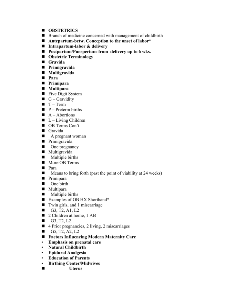
• • • • • OBSTETRICS Branch of medicine concerned with management of childbirth Antepartum-betw. Conception to the onset of labor* Intrapartum-labor & delivery Postpartum/Puerperium-from delivery up to 6 wks. Obstetric Terminology Gravida Primigravida Multigravida Para Primipara Multipara Five Digit System G – Gravidity T – Term P – Preterm births A – Abortions L – Living Children OB Terms Con’t Gravida A pregnant woman Primigravida One pregnancy Multigravida Multiple births More OB Terms Para Means to bring forth (past the point of viability at 24 weeks) Primipara One birth Multipara Multiple births Examples of OB HX Shorthand* Twin girls, and 1 miscarriage G3, T2, A1, L2 2 Children at home, 1 AB G3, T2, L2 4 Prior pregnancies, 2 living, 2 miscarriages G5, T2, A2, L2 Factors Influencing Modern Maternity Care Emphasis on prenatal care Natural Childbirth Epidural Analgesia Education of Parents Birthing Center/Midwives Uterus Has 3 layers: 1. Perimetrium 2. Myometrium 3. Endometrium Provides housing & nourishment Fertilized ovum implants there Myometrium* Muscular layer What tumors grow here? Fundus Upper thicker portion of uterus* Top portion where fallopian tubes connect Vagina 1. Birth canal 2. Organ of copulation 3. Excretes menstrual flow 4. Rugae 5. Bartholin’s Gland* Perineum Area bet. Posterior vaginal wall & anus* Provides muscular support for pelvic organs Structures that Support the Uterus Broad Ligament Round Ligament Uterosacral Ligament ***Support the uterus in it’s proper position*…… Terms Used in reference to the Female Pelvis A.Gynecoid Pelvis* 1. Wider than male pelvis Android pelvis is much narrower* 2. Contains & protects reproductive organs, bladder & rectum 3. Forms part of the birth canal 4. Larger than male pelvis 5. Formed by: coccyx, sacrum, hip bones(ilium,ishium & pubis) 6. 50% of woman have a gynecoid pelvis 2. False Pelvis The upper flaring part of pelvis* Supports growing uterus during pregnancy Offers landmarks for pelvic measurement Directs fetus toward True Pelvis 3. True Pelvis Formed by pubis(front), the ilia/ischia(sides) and sacrum/coccyx(behind)* The true pelvis has: Inletentrance from false pelvis* Cavitycurved area* Outletexit from True pelvis* The true and false pelvis are divided by an imaginary line called the Linea Terminalis or Pelvic Inlet Ischial spine refers to midpelvis* Hormonal Control/Menstrual Cycle Menstrual Cycle *usually 28 days *day 1menses begins *day 14ovulation occurs Fertilization Must occur w/I 48 hrs. of ovulation After fertilization6-8dys for zygote to travel for implantation Without fertilizationhormone levels drop, menstruation occurs. Hormonal Control FSHsecreted by ant. Pituitary* Stimulates graafian (ovarian) follicle in ovary Ovum matures in graafian follicle Estrogen secreted by graafian follicle as ovum matures Prepares uterus for pregnancy Thickens the endometrium Inhibits FSH Stimulates LH LH secreted by anterior pituitary Causes ovulation Transforms graafian follicle to corpus luteum Progesterone Secreted by corpus luteum Causes endometrium to thicken Essential in maintenance of pregnancy It inhibits uterine contractions Hormonal differences with &without fertilization With: 1.corpus luteum secretes progesterone & estrogen for 11-12 wks, then placenta takes over production of these hormones* 2.HCG is secreted HCG Human Chorionic Gonadotropic hormone Secreted by embroyonic cells tocontinue to stimulate the corpus luteum Without Fertilization: corpus luteum dies Estrogen & progesterone levels decrease Endometrial degeneration occurs & menstruation begins…. Conception & Implantation Conception • Takes place in fallopian tubes • Occurs after ovum & sperm unite There are 23 chromosomes each 46 chromosomes = Zygote 44 autosomes and 2 sex Male Y & X, Female X & X Each ejaculate has 200 – 400 million sperm cells Implantation o Zygote morula (mulberry) when it reaches the uterus o Morula Blastocyte when it enters uterus o Blastocyte implants into the endometrium & is now called the “Embryo” Peter, show transparancy Determination of Sex Determined by father’s sperm Female ovum has only “x” chromosome Male sperm has both “x” & “y” chromosome 2 Types of Multiple Births Monozygotic* A. Identical Twins (30%) 1.single ovum & sperm* 2.fertilized egg dev.2 embryos 3.usually 1 placenta (2 sacs) 4.always the same sex B. Fraternal Twins 1. Dizygotic Twins (70%) 2 ova & 2 sperm, both implant* 2. 2 placentas (separate or fused) w/ 2 sacs May/may not be same sex May/may not look alike Lab Tests to Determine Pregnancy Most based on presence of HCG in blood or urine HCG is present anywhere from 8-15 days after conception* Home test are 95% accurate Use first void in morning* Pregnancy tests HCG may show up as early as 7 – 8 days past ovulation Home kits will test positive around 14 days passed missed period E.D.C.- Estimated Date of Confinement Calculated by Nageles rule:* 1.count back 3 mos. From the 1st day of LMP 2.Add 7 days 9 months or 266 days Nagles calculations from LMP 6/25/07: - 3 months = 3/25, + 7 days = 4/1/08 2/25/08: - 3 months = 11/25, + 7 days = 12/2/08 01/01/08: - 3 months = 10/1, + 7 days = 10/08/2008 Determination of Pregnancy 3 Degrees of Certainty based on Symptoms a. Presumptive b. Probable c. Positive Presumptive Sx Amenorrhea Nausea & vomiting Frequent urination (1st & 3rd trimester)* Fatigue Breast changes Pigmentation Changes Chloasma, Linea Nigra, Periareola darkening Quickening(first fetal movement)* Change in abdomen shape & size* Chadwick’s Sign* Probable Signs & Sx + urine pregnancy test RIA test + Radioimmunoassy Goodell’s sign* Softening of cervix Hegar’s sign* Softening lower uterine segment Ballottement Feeling the fetal rebound when palpating uterus Positive Signs & Sx Fetal Heart Beat* faint @ 10-12 wks. With doppler ultrasound Distinct @ 18-20 wks. 120-160 beats/min.* Must take mother’s pulse at same time* X-ray visualization Interesting Stuff Tunic Souffle Swishing sound caused by pulsation of blood through unbilical cord. Rate is same as fetal HR Uterine (Placental) Souffle Swishing sound produced by maternal blood as it flows through large vessels of uterus. Rate is same as mother Maternal & Fetal Circulation A. Placenta Dark red circular organ Weighs ~ 1-2 lbs. A. B. C. D. E. F. G. 1. 2. 3. 1. Dev. From both embryonic & maternal tissue Totally formed and functioning by 12 wks.* Expands for 20 weeks, then grows only in thickness Maternal side (decidua basalis) Shiny Schultz Fetal side has chorionic villi Dirty Duncan Time Lines* 3rd month Placenta is complete 4th month Sex is differentiated 6th month Fingernails, eyebrows, eyelashes 8th month Embryo becomes a fetus and is viable 9th month Fat is stored Peter, show the movie”Nine Month Miracle Placental Functions: Provides for nutrition, excretion & respiration of fetus Secretes progesterone* Inhibits uterine contractions Secretes estrogen * Aids in lactation and fetal development Secretes HCG Acts as protective barrier Placental Transfer Exchange of nutrients, excrement & respiration* via osmosis and difusion There is NO intermixing of fetal & maternal blood ….* 2 separate systems B. Umbilical Cord Attaches fetus to placenta Contains 2 arteries & 1 vein (intertwined & covered by Wharton’s jelly)* 2 cm in diameter, 55 cm length C. Fetal Circulation Umbilical veincarries oxygenated blood & nutrients from placenta to fetus* 2. Umbilical arteriescarry waste products from fetus to placenta* Know….. 1.Ductus Venosus 2.Foramen Ovale* 3. Ductus Arteriosis Peter, show “Fetel Circulation” transparancy Fetal Circulation Umbilical vein passes through liver and the Ductus Venosus The blood goes to the RA Then shunted to left side via Foramen Ovale (RA to RV) Ductus Arteriosis ( PA and Aorta) The fetus does not use lungs, 02 is derived from placenta Fetal Circulation Con’t From Aorta, blood goes to head, trunk, extremities, internal iliac arteries to 2 umbilical arteries to placenta Fetal circulation at birth Lung function is established Umbilical arteries clot and turn into fibrous cords Umbilical vein forms round ligament of liver Foramen Ovale closes Ductus Venosus and Ductus Arteriosis shriven up Physiological Changes & Common Discomforts of Pregnancy A. Cardiovascular Blood volume up 30-40 %* Heart rate up BP remains unchanged Varicose veins* B. Respiratory • Rate is increased • Lung capacity is decreased C. Digestive Stomach & intestines displaced upwards Peristalsis slows constipation* Nauseau cau. By hormones* Heartburn due to reflux of stomach contents* Digestive con’t Constipation d/t decrease in peristalsis from progesterone. Teach to increase fluids and roughage* Heartburn d/t stomach contents flowing up esophagus. Teach not to recline for 30 min PP* Nausea d/t hormones. Teach to eat dry toast or crackers D. Endocrine Glands increase in size & activity Metabolic rate increases From increase in thyroxine Endocrine problems: Morning sickness, headaches, N&V, food cravings, breast enlargement E. Musculoskeletal Lordosis Pubic symphysis & sacroiliac joints become more pliable Pendulous abdomen strains M/S system F. Urinary • Kidney activity increases • Urinary frequency ( 1st and 3rd) trimesters * G. Integumentary Striae on abdomen, hips, thighs, & breasts Pigmented mask on face (chloasma)* Increase pigmentation abdomen (linea nigra) Prenatal/Antepartal Care GOAL: **Maximum physical & mental fitness of woman with an uncomplicated delivery & healthy newborn…** A. Routine Exams…. a. Q 4 wks 32 wks. b. Q 2 wks 36 wks. c. Weekly until 6 wks. Postpartum Exams include BP, wt, fundal ht, fetal heart rate. If mother is Rh (-) She will receive Rhogam at 28 weeks Erythroblastosis Fetalis Rh (+) fetus in Rh (-) mother Rh protein crosses placental barrier and invades mother Mother makes antibodies Antibodies cross over to fetus and destroys blood cells Treated with special gamma globulin called RhoGAM B. NUTRITIONAL NEEDS Diet based on Food Guide Pyramid Increase calories by 300 daily* Increase calcium 3 – 4 cups milk daily Meats ^zinc, iron and protein Folic acid supplements Reduces neural defects Nutrition con’t Desirable weight gain is 25 – 30 lbs* Calcium is important for bone growth and blood clotting of fetus Zinc helps the body with metabolism. It is an enzyme. Best source is meats.* Increase protein intake for fetus & mother Avoid empty calories Iodized salt Variety of foods No laxatives/enemas Use stool softeners instead Nutrition con’t Meats are best source of proteins and are good source of iron* Iron is stored by fetus for post birth, also used for new tissue growth. Iron is essential for production of Hgb and building and repair body tissues Increase fluids Increase vitamins Weight gain varies w/ weight of mother Do not diet Appetite ( Pica )* C. GENERAL HEALTH PRACTICES Left side lying Coping with stress Role/Relationship changes Self-perception/self concept changes Seat Belts Place beneath abdomen, over thigh and shoulder and between breasts Peter, transparency on lateral position D. TERATOGENIC FACTORS Teratogen is an environmental agent or factor that causes defects in fetus. Ex: Rubella, ETOH, smoking, drugs, dietary deficiencies Rubella* (-) rubella titer are not immune to rubella Will receive a rubella shot prior to discharge and told not to get pregnant for 3-6 months. Could cause fetal abnotmalies Smoking?* LBW Prematurity ETOH Fetal Alcohol Syndrome E. MINOR DISCOMFORTS 1. Morning sickness 2. Heartburn 3. Gingivitis 4. S.O.B. 5. Leg cramps 6. Varicose veins* 7. Vaginal discharge* Do not douche 8. Constipation 9. Supine hypotension 10. Backaches 11. Yellowish discharge from breasts* (colostrum) F. Danger Signals to Report Refer to page 657 Box 24-7 Number one danger signal? Vaginal bleeding Complications of Pregnancy 1. ABORTIONS A. Spontaneous B. Therapeutic Spontaneous Abortions • Threatened • Complete • Septic • • 1. 2. 3. Habitual Inevitable Incomplete Missed* Fetal demise but remainsin utero, may need D&E or oxytoxin Therapeutic Abortions Interruption of pregnancy for medical or social reasons. Some may be criminal or illegal Complications of Abortion Infection Hemorrhage Rh sensitization (occurs only with Rh- woman carrying an Rh+ fetus) 2. Premature Dilation of the Cervix “Incompetent Cervix” Caused by : *Previous cervical lacerations *Cervical or vaginal CA *Multiple D & C’s or biopsies Congenital (maternal exposure to DES (Diethylstilbestrol) TREATMENT: Cervical Cerclage 3. Ectopic Pregnancy Implantation occurs somewhere other than the uterus Other sites abdominal cavity, ovaries, ligaments or cervix 95% occur in fallopian tubes* Symptoms Sharp, localized, one-sided pain or pain referred to the shoulder Rigid and tender abdomen Slight vaginal bleeding Signs of hypovolemic shock Treatment Surgical treatment must be prompt* Salpingectomy Salpingostomy Methotrexate Destroys the rapidly dividing cells II. Maternal Disorders Affecting Pregnancy A. Hyperemesis Gravdiarum* Excessive vomiting Exact cause is unknown HCG is suspected Common nutrition-related discomforts of pregnancy Medical Management Meet nutritional needs Balance electrolytes with IV TPN used in severe cases Reintroduce solid foods slowly Prognosis is good B. Pregnancy Induced Hypertension ( PIH) Includes: Preeclampsia ( Mild or Severe) AND Eclampsia 1) 2) 3) 4) • • • • • Classic Signs….. Edema* Hypertension Proteinuria (albuminurea)* Signs generally occur after 20th wk of pregnancy Mild Preelampsia Few clinical symptoms BP of 140/90 Generalized edema of face, hands and ankles Weight increase, & 1-2+ albumin in urine Severe Preeclampsia Symptoms appear suddenly BP of 160/110 or greater Increased edema Dramatic increase in weight Increase urine albumin & decrease in urine amount Eclampsia Most severe form of PIH Characterized by seizures & coma Elevated BP, albuminuria and oliguria are common also Nursing Interventions I&O Daily weights* Monitor BP every 4 hrs. Quiet environment & bed rest* Magnesium Sulfate-> used to prevent convulsions*, & lower BP* TEACH…. Importance of compliance with therapy Importance of bedrest Continuous care is mandatory Nursing Alert : HELLP Syndrome H Hemolysis (destruction of RBC’s) EL Elevated Liver Enzymes LP Low Platelet Count C. Gestational Diabetes Diabetes during pregnancy Screened at 26-28 wks Ranges from diet controlledinsulin Most gestational diabetes return to normal after delivery Fetus will suffer from hyperglycemia in early pregnancy. Controlled glucose levels are very important Nursing Interventions Maintain normal blood glucose Teach how to administer insulin & regulate blood sugars Insulin may be required by both NIDDM and GDM Insulin will not cross placenta* III. DISORDERS AFFECTING THE FETUS 1. INFECTIONS T Toxoplasmosis* Kitty litter O Other R Rubella C Cytomegalovirus H Herpes 2. Rh Sensitization Less frequent today Rh+ proteins enter maternal circulation of Rh- mother & she now produces Rh+ antibodies. Antibodies destroy the fetus’ RBC’s causing “ Erythroblastosis Fetalis” Trmt Rhogam injections * Problems: Mom Rh (-), Dad and baby Rh(+)* 3. ABO Incompatability Mother A fetus B or AB Mother B fetus A or AB Mother O fetus A, B, or AB Usually see jaundice w/i 24 hrs Rx with phototherapy IV. PLACENTAL & AMNIOTIC DISORDERS 1. Placenta Previa Placenta partially or completely covers the cervical os* Complete with “total” coverage* Partial with incomplete coverage* Marginal Low implantation is the placenta situated in the lower uterine segment away from the internal os Predisposing Factors Numerous or closely spaced pregnancies Abnormalities in uterine structure Late fertilization Symptoms • • • • • a) b) c) d) e) f) Painless vaginal bleeding occurring after 20 wks. * The separation of placenta from uterine wall is painless Diagnosed by: Ultrasound TX : Bedrest/observation vag birth not preferred C-section preferred Fetal death may occur d/t hypoxia +/or prematurity* 2. Abruptio Placenta Abrupt premature separation of normally implanted placenta* Grave complication of late pregnancy Cause is unknown Predisposing Factors Hypertension Pre-eclampsia (PIH) Substance abuse Grand multipara Numerous abortions Symptoms are: Pain, dark red blood, tender uterus usually in last trimester* Strong, consistent contractions Rising fundus (uterine rigidity) may indicate retroplacental hemorrhage* Predisposing factors Hypertension Pre-eclampsia* Substance abuse Grand multip Numerous Abs Physical trauma Complications are: Fetal dangershypoxia,anemia, death* Bleeding into uterine muscle Loss of uterine tone DIC maternal death Treatment/Management is: Continuous fetal monitoring Monitor fundal height (marking to check for upward movement. Freq. VS If no fetal distress vag delivery In severe form C- section Read chap 27 to tie in RDS Assessing Fetal Status A. Amniocentesis B. Chorionic Villi Sampling C. Ultrasound Scanning D. Oxytocic Challenge Test (Stress Test) E. Nonstress Test (NST) Oxytoxic Challenge Test Evaluates fetal cardiac response to contractions Oxytoxin given until contractions are 3 in 10 minutes Late decels indicate utero-placental placement insufficient + outcome means that > 50% are late decels Non-Stress Test Fetal heart rate should go up with activity Fetal monitor placed on Mom Mom signals when she feels movement If FHR >, this is good If FHR does not change or <, this is bad ? O2 or think of placental insufficiency F. Fetal Biophysical Profile Non-Stress Test and ultra sound used together Monitors fetal breathing movements, fetal tone, brady moments and amniotic fluid volume G. Alphafetalprotein Done on mother Done 16 – 18 weeks of pregnancy Measures antigen carried by fetus Elevated levels indicate neural tube defects Low levels indicate Down’s Syndrome May also be measured in amniotic fluid Low levels = Down’s Syndrome High levels = Spina Bifida


