Neuromuscular Function
advertisement
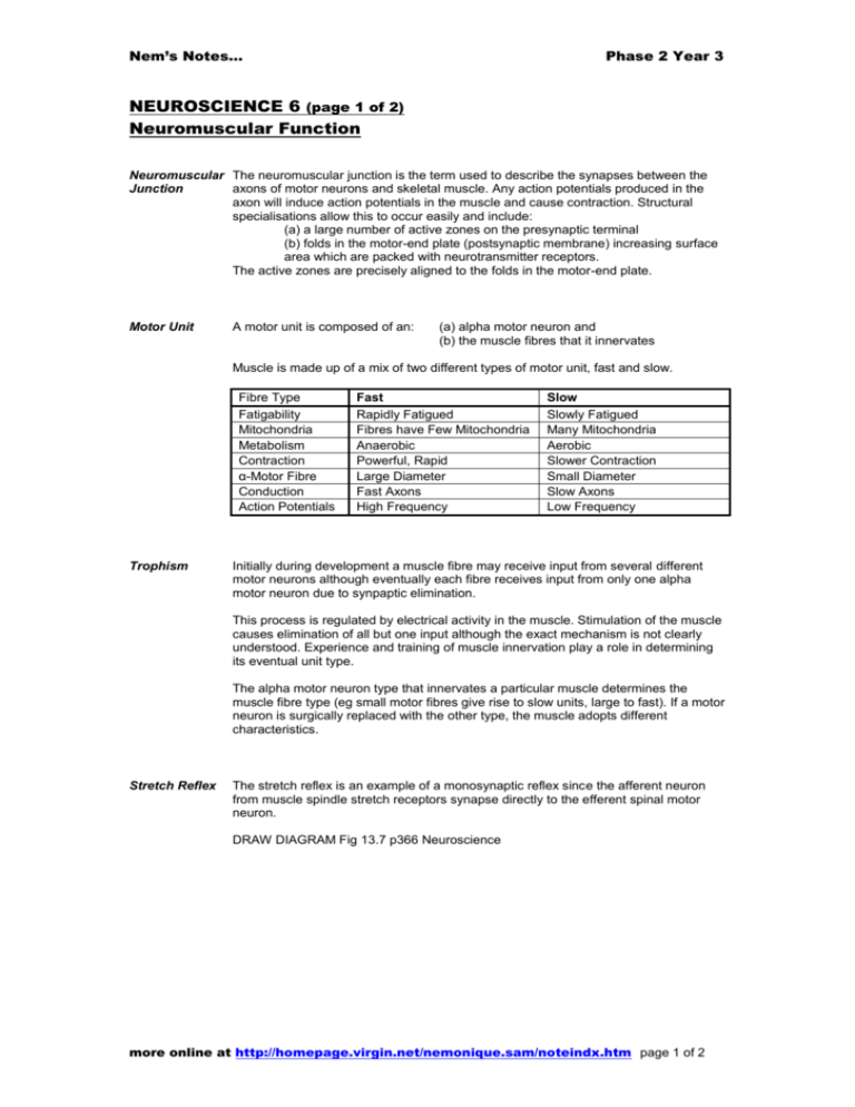
Nem’s Notes… Phase 2 Year 3 NEUROSCIENCE 6 (page 1 of 2) Neuromuscular Function Neuromuscular The neuromuscular junction is the term used to describe the synapses between the Junction axons of motor neurons and skeletal muscle. Any action potentials produced in the axon will induce action potentials in the muscle and cause contraction. Structural specialisations allow this to occur easily and include: (a) a large number of active zones on the presynaptic terminal (b) folds in the motor-end plate (postsynaptic membrane) increasing surface area which are packed with neurotransmitter receptors. The active zones are precisely aligned to the folds in the motor-end plate. Motor Unit A motor unit is composed of an: (a) alpha motor neuron and (b) the muscle fibres that it innervates Muscle is made up of a mix of two different types of motor unit, fast and slow. Fibre Type Fatigability Mitochondria Metabolism Contraction α-Motor Fibre Conduction Action Potentials Trophism Fast Rapidly Fatigued Fibres have Few Mitochondria Anaerobic Powerful, Rapid Large Diameter Fast Axons High Frequency Slow Slowly Fatigued Many Mitochondria Aerobic Slower Contraction Small Diameter Slow Axons Low Frequency Initially during development a muscle fibre may receive input from several different motor neurons although eventually each fibre receives input from only one alpha motor neuron due to synpaptic elimination. This process is regulated by electrical activity in the muscle. Stimulation of the muscle causes elimination of all but one input although the exact mechanism is not clearly understood. Experience and training of muscle innervation play a role in determining its eventual unit type. The alpha motor neuron type that innervates a particular muscle determines the muscle fibre type (eg small motor fibres give rise to slow units, large to fast). If a motor neuron is surgically replaced with the other type, the muscle adopts different characteristics. Stretch Reflex The stretch reflex is an example of a monosynaptic reflex since the afferent neuron from muscle spindle stretch receptors synapse directly to the efferent spinal motor neuron. DRAW DIAGRAM Fig 13.7 p366 Neuroscience more online at http://homepage.virgin.net/nemonique.sam/noteindx.htm page 1 of 2 Nem’s Notes… Phase 2 Year 3 NEUROSCIENCE 6 (page 2 of 2) Neuromuscular Function Flexor Reflex The flexor reflex is an example of a polysynaptic reflex. This is a complex reflex arc which is used to withdraw limbs from aversive stimuli. A single nociceptive input causes activation of excitatory interneurons which cause contraction of all flexor muscles in the affected limb. At the same time a crossed extensor reflex occurs in the contralateral limb causing contraction of all the extensor muscles. This provides postural support during the reflex. DRAW DIAGRAM Fig 4.9 p54 CRASH COURSE Pathological Reflexes Hyperreflexia is an abnormal exaggerated reflex response to a stimulus. It is often a sign of upper motor neuron dysfunction. Upper motor neurons are those from the cortex or brainstem connecting to the interneurons in the anterior horn of the spinal cord. Hyporeflexia is an absence of, or diminished reflex to, a stimulus and can be due to muscle fibre denervation and subsequent muscle atrophy, myopathies or lower motor neuron dysfunction. Lower motor neurons are those from the spine to the muscle. Trauma to the spinal cord can also affect reflexes due to disruption of the spinal arc. more online at http://homepage.virgin.net/nemonique.sam/noteindx.htm page 2 of 2

