13 and 14
advertisement
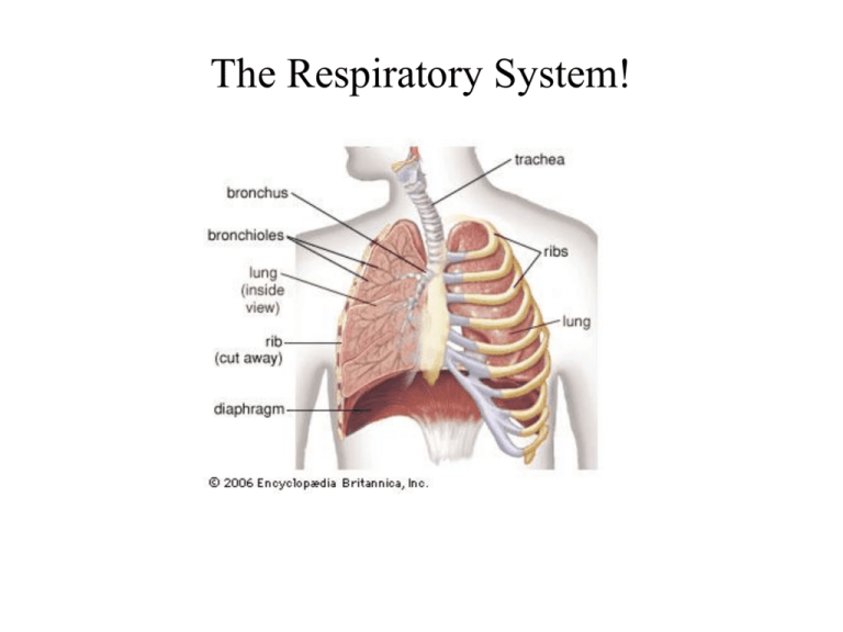
The Respiratory System! U of Mich Med site RESPIRATORY SYSTEM The function of the respiratory system is to provide O2 and to remove CO2 from the blood Segments Conducting Portion consisting of the nasal cavity, larynx, trachea, bronchi and bronchioles Transitional Portion formed by the respiratory bronchioles Respiratory Portion formed by the alveolar duct, alveolar sac and the lung alveoli Function Maintain an open passageway for air passage, condition air headed for lungs (by increasing humidity and temperature) Combination of conducting and respiratory functions: Keep airways open for easy air passage, condition air headed for lungs (by increasing humidity and temperature) and start of gas exchange. Exchange of O2 and CO2 between air and blood Features Presence of cartilage and smooth muscle cells in airways Presence of smooth muscle cells, but NO cartilage No cartilage or smooth muscle cells, presence of alveolar sacs. GENERAL PLAN OF THE CONDUCTING PORTION (TRACHEA) Muscle Cartilage Submucosa Epithelium* Lamina Propria Adventitia * Pseudo stratified columnar ciliated epithelium with goblet cells Mucosa GENERAL PLAN OF THE CONDUCTING PORTION (TRACHEA) Pseudo stratified columnar ciliated epithelium with goblet cells Mucosa Lamina Propria ( loose C.T. , elastic fibers, capillaries) Submucosa C.T. and Glands Cartilage Muscle In the trachea, hyaline cartilage Adventitia Dense irregular C.T. Trachealis Muscle (smooth muscle) Acinar Glands (in submucosa) Cartilage Epithelium Poor stain packed with glycoproteins Nucleus at base Sticky secretion Acidophilic Central, round nucleus Watery fluid Serous Acinus Mucous Acinus PSEUDO STRATIFIED COLUMNAR CILIATED EPITHELIUM Ciliated Cells Goblet Cell Brush Cell BM * Epinephrine, Serotonin ** Squamous Metaplasia Small Granule Short Cell (stem ?) Cell (Argentaffin *) (250-300 cell cilia) Microtubule Core Basal Body Z.A. + Actins (Microfilaments) Centrioles (Nucleation Center) Desmosome + Intermediate Filaments Microtubules Cytoskeleton Microfilaments (Actins) 10 nm Intermediate Filaments 15 nm * Microtubules 25 nm * Cytokeratin, Vimentin, Desmin, Neurofilaments MTs are essential for a broad range of cell functions, most notab Mitosis – segregation of chromosomes Transport – molecular motors (Kinesins and Dynein) Cell Motility – Cilia and Flagella Cell shape and Polarity (eg. Axon in neurons) Positioning of membrane enclosed organelles (golgi and ER) Cilium Microtubule core a( xoneme) AXONEME 9 pairs of microtubules Basal Body (B.B.) 1 central pair Centrioles PM Basal Body B.B. Microtubules Each centriole of the pair shown in the centrosome has the same structure of the basal body. Each centriole (or basal body) is a cylinder, 0.2 m wide X 0.4 m long Cilium Axoneme 24nm Cross section Basal body Basal Body (Centriole) 9 Dynein Arm Radial 8 Spokes B Central Pair A 7 6 5 Outer Doublet Tektin Central Sheath Nexin bridge Triplets The stable MTs in the cilium are arranged in a characteristic pattern of nine outer doublet MTs – A and B. One MT (A) in each doublet is complete while the other (B) contains only 11 protofilaments. They are fused and share a common wall. Composed of 9 sets of triplet MTs. Each MT triplet contains one complete MT (A tubule) and 2 attached incomplete MTs (B & C tubules) which share walls with the adjacent MTs. Other proteins from crosslink between these triplet Protein Linker MTs Ciliary movement the axoneme is mediated by axonemal and ciliary dynein. The tail region of the dynein attaches to the A tubule while the head region with the motor interacts with the B tubule of the adjacent doublet. B.B. PM M Anatomy of Respiration Trachea Conducting Pulmonary Bronchi Regular Bronchioles Transitional Respiratory Terminal Bronchioles Respiratory Bronchioles Alveolar Ducts Alveoli Alveolar sacs GENERAL PLAN OF THE CONDUCTING PORTION (TRACHEA) Muscle Cartilage Submucosa Epithelium* Lamina Propria Adventitia * Pseudo stratified columnar ciliated epithelium with goblet cells Mucosa EP LP HC EP LP SM SM Adventitia HC Glands Note: As you move down the airway - less goblet cells, - less mucous acini, more serious acini - cartilage pieces - no c-rings 17-7 SM SM LP EP LP Adventitia * asthma EP EP SM EP LP LP SM * Clara cells EP* SM EP* SM * Clara cells Arrows indicate knobs of mucosa Respiratory Bronchiole Alveolar Duct Alveoli Septum Alveolar Sac Bronchial Tree - Fig. 17-16 Fig.17-19 Pneumocyte II Macrophage Pneumocyte I Alveolar Lumen Septum Endothelium (Capillary) Septum THE BLOOD-AIR BARRIER Surfactant BM H2CO3 O2 CO2 H2CO3 + Carbonic Anhydrase = CO2 + H2O MVB (MVB) The lamellar bodies contain the surfactant proteins A,B, C and D and phospholipids, primarily lecithin, which help to reduce the surface tension of the pneumocytes I.
