American College of Radiology ACR Appropriateness Criteria®
advertisement
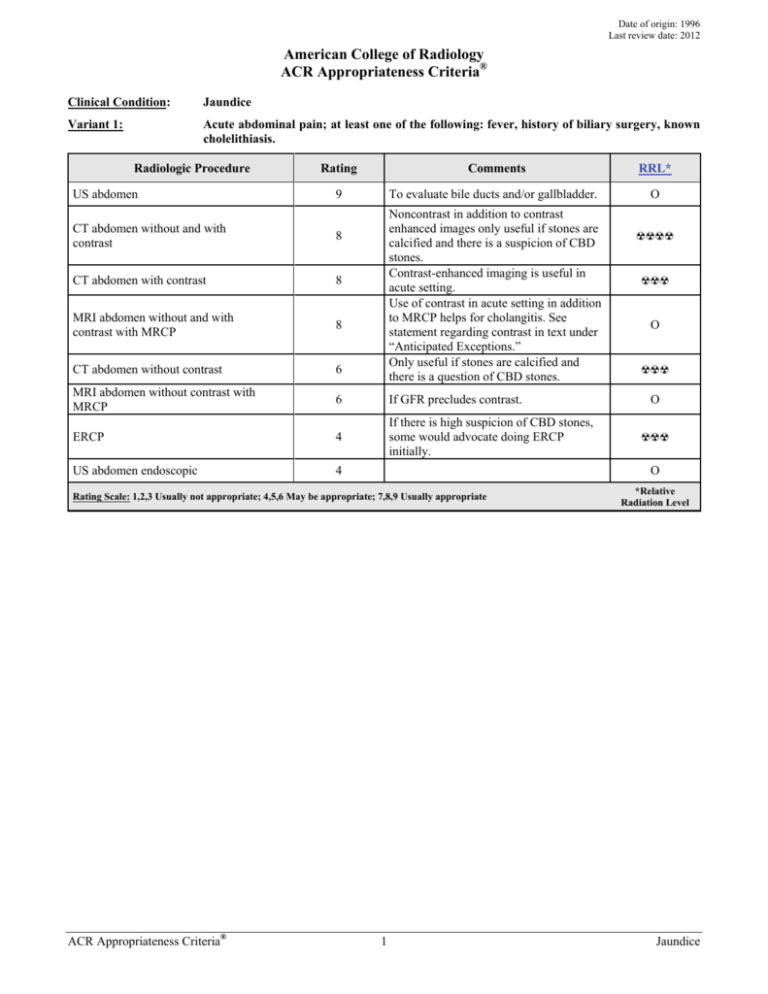
Date of origin: 1996 Last review date: 2012 American College of Radiology ACR Appropriateness Criteria® Clinical Condition: Jaundice Variant 1: Acute abdominal pain; at least one of the following: fever, history of biliary surgery, known cholelithiasis. Radiologic Procedure US abdomen Rating Comments RRL* 9 To evaluate bile ducts and/or gallbladder. O Noncontrast in addition to contrast enhanced images only useful if stones are calcified and there is a suspicion of CBD stones. Contrast-enhanced imaging is useful in acute setting. Use of contrast in acute setting in addition to MRCP helps for cholangitis. See statement regarding contrast in text under “Anticipated Exceptions.” Only useful if stones are calcified and there is a question of CBD stones. CT abdomen without and with contrast 8 CT abdomen with contrast 8 MRI abdomen without and with contrast with MRCP 8 CT abdomen without contrast 6 MRI abdomen without contrast with MRCP 6 If GFR precludes contrast. ERCP 4 If there is high suspicion of CBD stones, some would advocate doing ERCP initially. US abdomen endoscopic 4 ☢☢☢ O ☢☢☢ O ☢☢☢ O Rating Scale: 1,2,3 Usually not appropriate; 4,5,6 May be appropriate; 7,8,9 Usually appropriate ACR Appropriateness Criteria® ☢☢☢☢ 1 *Relative Radiation Level Jaundice Clinical Condition: Jaundice Variant 2: Painless; one or more of the following: weight loss, fatigue, anorexia, duration of symptoms greater than 3 months. Patient otherwise healthy. Radiologic Procedure Rating CT abdomen with contrast 8 US abdomen 8 MRI abdomen without and with contrast with MRCP 8 CT abdomen without and with contrast MRI abdomen without contrast with MRCP Comments Biphasic arterial and portal venous phase may be useful and provides greater dose reduction than CT without and with contrast. If there is high pretest probability of obstruction due to malignancy, can be alternative to US. To evaluate bile ducts and/or gallbladder. May be the first-line test to evaluate for obstruction. If there is high pretest probability of obstruction due to malignancy, can be alternative to US and may provide roadmap for subsequent therapeutic intervention. See statement regarding contrast in text under “Anticipated Exceptions.” 7 6 CT abdomen without contrast 5 US abdomen endoscopic 5 If GFR precludes contrast. Not as an initial test. Would do imaging study first. May help to evaluate for dilated duct when GFR is low and patient is not able to have MRI. May be appropriate as a therapeutic and diagnostic intervention for mass at the ampulla and periampullary region and may further staging. Rating Scale: 1,2,3 Usually not appropriate; 4,5,6 May be appropriate; 7,8,9 Usually appropriate ACR Appropriateness Criteria® ☢☢☢ O O ☢☢☢☢ 7 ERCP RRL* 2 O ☢☢☢ ☢☢☢ O *Relative Radiation Level Jaundice Clinical Condition: Jaundice Variant 3: Clinical condition and laboratory examination make mechanical obstruction unlikely. Radiologic Procedure Rating US abdomen 8 MRI abdomen without and with contrast with MRCP 8 CT abdomen with contrast 7 MRI abdomen without contrast with MRCP 6 CT abdomen without and with contrast 6 CT abdomen without contrast 5 ERCP 3 US abdomen endoscopic 3 Comments To evaluate liver for echogenicity, nodularity, stigmata of cirrhosis. Initial test. If US is equivocal or does not answer the clinical question. Excludes biliary source of jaundice. Can visualize contour of liver, assess for liver iron and fat. Additional CE images may enable characterization of mass in cirrhotic livers. See statement regarding contrast in text under “Anticipated Exceptions.” Multiphase liver. Excludes biliary source of jaundice. Can visualize contour of liver, assess for liver iron and fat. Noncontrast CT can help to confirm fatty liver in cases of NAFLD; contrastenhanced multiphase CT can enable lesion characterization in cirrhotic livers. Useful for assessing liver contour and NAFLD but does not exclude lesions. Not as an initial test. Would do imaging study first. May be useful only if imaging study does not yield a diagnosis. Rating Scale: 1,2,3 Usually not appropriate; 4,5,6 May be appropriate; 7,8,9 Usually appropriate ACR Appropriateness Criteria® 3 RRL* O O ☢☢☢ O ☢☢☢☢ ☢☢☢ ☢☢☢ O *Relative Radiation Level Jaundice JAUNDICE Expert Panel on Gastrointestinal Imaging: Tasneem Lalani, MD1; Corey A. Couto, MD2; Max P. Rosen, MD, MPH3; Mark E. Baker, MD4; Michael A. Blake, MB, BCh5; Brooks D. Cash, MD6; Jeff L. Fidler, MD7; Frederick L. Greene, MD8; Nicole M. Hindman, MD9; Douglas S. Katz, MD10; Harmeet Kaur, MD11; Frank H. Miller, MD12; Aliya Qayyum, MD13; William C. Small, MD, PhD14; Gary S. Sudakoff, MD15; Vahid Yaghmai, MD, MS16; Gail M. Yarmish, MD17; Judy Yee, MD.18 Summary of Literature Review Background/Introduction One of the difficulties in determining a rational imaging strategy to evaluate jaundiced patients stems from the fact that jaundice is a clinical finding, not a single disease entity. The causes of nonhemolytic jaundice can be divided into two distinct categories: intrahepatic biliary stasis (hepatocellular jaundice) and mechanical biliary obstruction. Because imaging plays little useful role in the evaluation of intrahepatic biliary stasis, the first task of the clinician caring for the jaundiced patient is to determine if jaundice is caused by bile duct obstruction. Several studies have shown that this distinction can be made in approximately 85% of patients using only clinical findings (age, nutritional status, pain, systemic symptoms, stigmata of liver disease, palpable liver or gallbladder) and simple biochemical tests [1]. Patients with a high pretest probability of nonobstructive jaundice usually have either diffuse hepatocellular disease (eg, cirrhosis, hepatitis), or, more rarely, inability of the liver to handle a bilirubin load (eg, hemolytic anemia), or a metabolic deficiency (Gilbert’s disease). These patients need no imaging studies; biopsy is usually the next step as clinically indicated. Obstructive jaundice is jaundice resulting from obstruction to the flow of bile from the liver to the duodenum. In adults, extrahepatic (mechanical) obstruction accounts for 40% of patients presenting with jaundice as the primary symptom [1], and this likelihood increases with advancing age. The most common causes of obstructive jaundice in the United States are neoplasms of the pancreas, ampulla of Vater or biliary tract, choledocholithiasis, pancreatitis, and iatrogenic strictures of the biliary tree. Other less common causes include tumors metastatic to the biliary epithelium, sclerosing cholangitis, hepatic tumors adjacent to the hilum, perihepatic lymphadenopathy, and other causes of cholangitis. Imaging Methods The methods used in evaluating the jaundiced patient currently include ultrasound (US), computed tomography (CT), magnetic resonance cholangiopancreatography (MRCP), and endoscopic retrograde cholangiopancreatography (ERCP). These examinations are effective to varying degrees for assessing both the cause and the site of obstruction; ERCP also can relieve the obstruction in a significant portion of cases [2-4]. Endoscopic US (EUS) can be used as an adjunct examination to ERCP in cases of common bile duct (CBD) obstruction and can be used to determine whether the obstruction is from mass or stone [5-9]. The literature is replete with articles confirming the usefulness of all of these methods, but comparative studies have rarely considered the effect of factors that may influence the validity of their conclusions. Among these factors are the prevalence of extrahepatic obstruction in the population studied, the various causes of obstruction (case mix) in the series (often a function of institutional bias), and the frequency of uninterpretable results or unsuccessful studies. These factors influence the apparent differences in efficacy [10]. In designing appropriateness criteria, therefore, we have chosen to consider strategies in terms of the pretest probability that jaundice is due to either mechanical obstruction or parenchymal liver disease. 1 Principal Author and Panel Vice-chair, Inland Imaging Associates and University of Washington, Seattle, Washington. 2Research Author, Beth Israel Deaconess Medical Center, Boston, Massachusetts. 3Panel Chair, Beth Israel Deaconess Medical Center, Boston, Massachusetts. 4Cleveland Clinic, Cleveland, Ohio. 5Massachusetts General Hospital, Boston, Massachusetts. 6Walter Reed National Military Medical Center, Bethesda, Maryland, American Gastroenterological Association. 7Mayo Clinic, Rochester, Minnesota. 8Carolinas Medical Center, Charlotte, North Carolina, American College of Surgeons. 9 New York University Medical Center, New York, New York. 10Winthrop University Hospital, Mineola, New York. 11MD Anderson Cancer Center, Houston, Texas. 12Northwestern University Feinberg School of Medicine/NMH, Chicago, Illinois. 13University of California San Francisco, San Francisco, California. 14Emory University, Atlanta, Georgia. 15Medical College of Wisconsin, Milwaukee, Wisconsin. 16Northwestern University, Chicago, Illinois. 17 Staten Island University Hospital, Staten Island, New York. 18University of California San Francisco, San Francisco, California. The American College of Radiology seeks and encourages collaboration with other organizations on the development of the ACR Appropriateness Criteria through society representation on expert panels. Participation by representatives from collaborating societies on the expert panel does not necessarily imply individual or society endorsement of the final document. Reprint requests to: Department of Quality & Safety, American College of Radiology, 1891 Preston White Drive, Reston, VA 20191-4397. ACR Appropriateness Criteria® 4 Jaundice It must be remembered that the results of any given imaging method strongly depend on the population studied and the expertise of the examiners. For this reason, local conditions and expertise should properly influence the method by which jaundiced patients are evaluated. Radiographs Radiographs rarely provide any information on the site or the cause of obstruction and have no place in the evaluation of the jaundiced patient. Ultrasound US is the least invasive and lowest-cost imaging technique available for evaluating obstructive jaundice. US is used to determine the presence of obstructive jaundice by depicting dilated bile ducts, with sensitivity of 55%95% and specificity of 71%-96% [2]. False-negative studies are due to two factors: inability to visualize the extrahepatic biliary tree (often because of interposed bowel gas), and the absence of biliary dilation in the presence of obstruction. US is less effective than CT or MRCP for determining the site and the cause of obstruction [2,11]. Computed Tomography CT is slightly more sensitive (74%-96%) and specific (90%-94%) than US for detecting biliary obstruction; in addition, the ability to determine the site and the cause of obstruction is greater with CT than with US. Recent articles claim that its sensitivity for obstruction is >90%, especially given the advent of multidetector CT (MDCT) and image reformations [12,13]. CT is strongly recommended as the primary modality for evaluating patients with suspected malignant biliary obstruction, both for diagnosis and for staging [12,14]. CT cholangiopancreatography generated by slab volume imaging with minimum-intensity projections (MinIp) and curved planar reformations may be useful for preintervention planning [14,15]. Recent studies also examine the utility of CT for predicting tumor extension and potential resectability [14,16,17]. Magnetic Resonance Imaging Magnetic resonance imaging (MRI) can demonstrate both the site and cause of biliary obstruction. MR cholangiography has been shown to be useful in depicting the three-dimensional anatomy of the biliary and pancreatic ducts. For detection of ductal calculi, MRCP is the most sensitive of the noninvasive techniques [1721]. The use of MRCP may decrease the number of ERCP examinations obtained prior to elective cholecystectomy [21,22]. More recent studies have recommended MRCP as the preferred test in patients with a high likelihood of choledocholithiasis. MRCP is valuable in the clinical situation of failed ERCP [21,23] and in patients with hilar biliary obstruction due to ductal tumor or periductal compression [17,19,24-27]. MRCP is the test of choice in pregnant women with known or suspected pancreaticobiliary disease due to lack of nonionizing radiation. [22]. Endoscopic Retrograde Cholangiopancreatography ERCP is the most common invasive diagnostic biliary procedure and has evolved gradually from its initial role as a diagnostic tool. Due to significant advances in cross-sectional imaging, in particular the advent of MRCP, ERCP currently has an almost exclusively therapeutic role [28]. Due to its inherent risks, costs, and invasive nature, ERCP should be indicated only for therapeutic reasons or when it can alter patient management. In addition, the procedure carries a potentially severe morbidity of up to 10%, most commonly pancreatitis, and a 0.4% mortality rate. These factors need to be weighed against the potential benefits of ERCP [3,23,29]. The main indication for ERCP remains management of CBD stones, which can be cleared in 80%-95% of cases [3,30]. Some also advocate ERCP in the management of severe acute biliary pancreatitis, along with cases of more mild disease complicated by one of the following: presence of gallstones plus high operative risk, absence of gallstones or prior cholecystectomy, or pregnancy. ERCP also remains the standard for stent placement in cases of obstructive jaundice. When deployed for distal CBD strictures, stenting via ERCP is successful in more than 90% of cases [31]. Stent deployment can be more complicated in cases of hilar cholangiocarcinoma, when placement of more than one stent could be necessary to achieve adequate drainage of the biliary tree [32]. There remains a limited role for diagnostic ERCP, essentially restricted to tissue sampling from biliary or pancreatic lesions, sphincter of Oddi manometry, and diagnostic pancreatoscopy or cholangioscopy [29]. Endoscopic Ultrasound EUS, an adjunct procedure to ERCP, can be used to detect small distal biliary ductal calculi, for local staging of periampullary neoplasm, and for guided fine-needle aspiration (FNA) biopsy [5,7,11,33]. The sensitivity, specificity, positive predicative value, and accuracies of EUS FNA biopsy for tumor are 84.6%, 100%, 100%, and ACR Appropriateness Criteria® 5 Jaundice 87.8%, respectively [7]. Recent literature also supports the use of EUS as an adjunct in evaluating potential duct strictures as well as early-stage tumors in the nonicteric stage [5,8,9]. Clinical Categories To determine the appropriateness of any imaging test, it is necessary to consider the general clinical category to which the patient belongs. The major categories are 1) high likelihood of a mechanical obstruction, 2) low likelihood of mechanical obstruction, and 3) indeterminate. For situations in which the preimaging probability of obstruction is high, it is also appropriate to consider a second question: whether the obstruction is likely to be benign or malignant. Variant 1: High Likelihood of Benign Biliary Obstruction Patients in this category present with jaundice and acute abdominal pain. There may be a prior history of gallstones documented by US or of prior biliary surgery. US is a readily available and inexpensive method for detecting dilated intrahepatic bile ducts and the common hepatic duct at the hepatic hilum. Biliary ductal calculi are not detected with the same sensitivity as gallbladder calculi [21,34]. In fact, the sensitivity for detection of CBD stones on US is 30% [21,34], as the subhepatic common duct may not be visible due to overlaying bowel gas. In addition, intrahepatic bile ducts may not be dilated in the early phase of acute obstruction or in patients with partial obstruction. Sensitivity of detection can be increased to 70%-86% by combining tissue harmonic imaging, elevated bilirubin, age >55 years, and CBD dilatation between 6-10 mm [21,35]. Despite recognized limitations, US is recommended as the initial diagnostic test in patients with suspected calculus obstruction of the common duct. In patients with acute biliary obstruction and suspected complicating conditions such as cholangitis, cholecystitis, or pancreatitis not well evaluated by US, a preintravenous and postintravenous contrast-enhanced abdominal CT study is useful in defining the level of obstruction, likely cause, and coexistent complications. CT can be used to detect partially calcified biliary calculi, but is insensitive for detecting bilirubinate or cholesterol calculi [13,15]. Isotropic data routinely obtained with current multislice technology can be reconstructed using narrow collimation, and smaller reconstruction intervals allow for better visualization of the calculi [13,15]. MRCP and ERCP are sensitive for detecting biliary ductal calculi [21,23]. The use of MRCP will improve the therapeutic yield of ERCP; however, MRCP has diminishing sensitivity with decreasing stone size <6 mm [36,37]. For these small stones EUS is most useful, as decreasing stone size does not hamper its performance, and complications with diagnostic EUS are rare [38]. Therapeutic endoscopic intervention including sphincterotomy may be curative, but it has associated morbidity of up to 10% due to risk of iatrogenic pancreatitis [23,29]. In patients with previous gastroenteric anastomoses, MRCP is recommended as the technique of choice to evaluate the extrahepatic biliary ductal system. In patients with suspected sclerosing cholangitis or biliary stricture, MRCP is the preferred imaging modality, avoiding the possibility of suppurative cholangitis that may be induced by endoscopic catheter manipulation of an obstructed biliary system [23]. MRCP findings may guide directed approaches such as ERCP with brushing, percutaneous transhepatic biliary stenting, or reconstructive surgery [17,19,20,23,25,39]. Variant 2: High Likelihood of Malignant Biliary Obstruction Patients typically present with insidious development of jaundice and associated constitutional symptoms (weight loss, fatigue, etc). Mechanical biliary obstruction can be confirmed by US. Malignant obstruction is most commonly due to pancreatic carcinoma but may be secondary to cholangiocarcinoma of either the proximal or distal duct or to periductal nodal compression. 64-slice MDCT using MinIp and multiplanar reconstructions (MPRs) shows excellent spatial resolution and accuracy for staging of biliary malignancies and helps differentiate benign from malignant strictures [12,14,16,40,41]. Reported sensitivity, specificity, and accuracy are 95%, 93.35% and 88.5%, respectively [12]. Important information in tumor staging includes tumoral involvement of the biliary confluence, invasion of the superior mesenteric and portal vein, peripancreatic tumor extension, regional adenopathy, and hepatic metastases [42]. Biphasic CT of the abdomen with pancreatic and portal venous phase imaging through the liver, biliary tree, and pancreas is the standard protocol for staging of pancreaticobiliary malignancies. However, improved z-axis resolution, isotropic data sets, and improved multiplanar capability must be balanced with the increased radiation dose to the patient in the acquisition of multiple phases [16]. ACR Appropriateness Criteria® 6 Jaundice MRI and MRCP are also accurate for tumor detection and staging. For example, accuracy rates for MRI with MRCP and MDCT are similar: 90.7% versus 85.1% for bilateral secondary biliary confluence involvement and 87% for both in detecting intrapancreatic CBD involvement in bile duct malignancies [17]. ERCP is invasive and more expensive than CT or MRI and has equivalent or greater sensitivity for tumor detection, particularly for ampullary carcinoma, but it does not provide staging information for operability [11]. Tissue diagnosis can be obtained by endoscopically directed brushing or guided US with FNA [3,5,6,8,11]. Additionally, in patients with suspected malignant biliary obstruction and negative or equivocal CT or MRI examinations, ERCP with EUS may provide an imaging and cytologic diagnosis (FNA) [5,9]. Endoscopic or percutaneous transhepatic biliary drainage is appropriate for patients who are not candidates for surgery and may even be useful in operative candidates for whom there is a delay to definitive surgical resection. Standard ERCP is sufficient in 90%-95% of patients who require biliary decompression. Factors that contribute to ERCP failure include gastric outlet or duodenal obstruction due to tumor invasion, or altered anatomy from diverticula or prior surgery. Percutaneous transhepatic cholangiography (PTC) can be used for decompression but requires external drainage, which can be a source of biliary infection. EUS-guided biliary drainage offers less morbidity as no external drainage is required, and it is increasingly used as a replacement for PTC [6]. In patients without pancreaticobiliary malignancy, periductal nodal disease can result in obstruction. Here, a diagnosis of lymphoma or metastatic adenopathy can be obtained with either image-guided biopsy or EUS. Variant 3: Low Likelihood of Mechanical Biliary Obstruction In situations where the pretest probability of mechanical biliary obstruction is low, either US or MRCP can be used as first-line imaging techniques because of their availability, absence of ionizing radiation, and low complication rates. US is less expensive though less definitive, especially for distal CBD and for ampullary and pancreatic visualization. When most of the abdominal organs need to be assessed, either CT or MRI can be used, though CT more reliably displays all abdominal anatomy. When CT evaluation is compromised (eg, in patients unable to receive iodinated intravenous contrast material), the combination of MRI and MRCP is a reliable alternative to exclude parenchymal cause for jaundice and to confirm that the pancreaticobiliary system is unobstructed. When imaging does not yield a cause for jaundice (ie, there is no obstruction and no parenchymal process to explain jaundice), liver dysfunction or infiltrative process must be excluded, and liver biopsy will be the most effective next step in diagnosis. Special Considerations The above situations address the initial workup of the jaundiced patient. It is assumed that once the initial workup has been performed and etiology of the obstruction ascertained, those patients with a mechanical obstruction will be triaged into endoscopic or percutaneous biliary drainage to alleviate the obstruction. (See the ACR Appropriateness Criteria® on “Radiologic Management of Benign and Malignant Biliary Obstruction.”) For those patients without mechanical obstruction, biopsy will help to elucidate the underlying process, and subsequent diagnosis will dictate follow-up. Summary The diagnostic approach for adults presenting with jaundice depends to a large extent on: clinical symptoms of pain, prior history of gallstones, acuity and/or duration of jaundice, and associated symptoms. The first objective of imaging is to decide whether there is mechanical obstruction. US as a first-line modality helps to confirm bile duct dilatation if present to assess for the presence or absence of stones, and to direct the second-line test. If a mechanical cause is suspected and there is associated right upper quadrant pain and a history of stones, MRI and MRCP may be the second modality performed. If there is mechanical obstruction but no stone disease and high suspicion of malignancy, then biphasic pancreatic CT with thin reconstructions can help define the point of obstruction, assess for resectability, and stage for metastatic disease. If no mechanical cause for jaundice is identified by the first-line modality of US, it is important to exclude infiltrative disease before performing invasive testing such as liver biopsy. Here, the superior tissue characterization offered by MRI may help further the diagnosis and may help direct biopsy. ACR Appropriateness Criteria® 7 Jaundice The availability of each modality and the expertise with which it is offered, institutional and referring physician preference, inherent patient characteristics (renal insufficiency, claustrophobia, implanted devices, ability to lay still), and radiation dose will also affect the choice. CT is readily available even in small rural hospitals but comes with inherent dose to patient. MRI may not be as readily available and may be difficult to obtain emergently, but it can be an alternative to CT when radiation dose is a consideration, such as in a younger or pregnant patient. All these modalities will have similar sensitivity, specificity and accuracy for depicting malignant disease, though MRI will be slightly superior for stone disease. Anticipated Exceptions Nephrogenic systemic fibrosis (NSF) is a disorder with a scleroderma-like presentation and a spectrum of manifestations that can range from limited clinical sequelae to fatality. It appears to be related to both underlying severe renal dysfunction and the administration of gadolinium-based contrast agents. It has occurred primarily in patients on dialysis, rarely in patients with very limited glomerular filtration rate (GFR) (ie, <30 mL/min/1.73m2), and almost never in other patients. There is growing literature regarding NSF. Although some controversy and lack of clarity remain, there is a consensus that it is advisable to avoid all gadolinium-based contrast agents in dialysis-dependent patients unless the possible benefits clearly outweigh the risk, and to limit the type and amount in patients with estimated GFR rates <30 mL/min/1.73m2. For more information, please see the ACR Manual on Contrast Media [43]. Relative Radiation Level Information Potential adverse health effects associated with radiation exposure are an important factor to consider when selecting the appropriate imaging procedure. Because there is a wide range of radiation exposures associated with different diagnostic procedures, a relative radiation level (RRL) indication has been included for each imaging examination. The RRLs are based on effective dose, which is a radiation dose quantity that is used to estimate population total radiation risk associated with an imaging procedure. Patients in the pediatric age group are at inherently higher risk from exposure, both because of organ sensitivity and longer life expectancy (relevant to the long latency that appears to accompany radiation exposure). For these reasons, the RRL dose estimate ranges for pediatric examinations are lower as compared to those specified for adults (see Table below). Additional information regarding radiation dose assessment for imaging examinations can be found in the ACR Appropriateness Criteria® Radiation Dose Assessment Introduction document. Relative Radiation Level Designations Relative Adult Effective Pediatric Radiation Dose Estimate Effective Dose Level* Range Estimate Range O 0 mSv 0 mSv <0.1 mSv <0.03 mSv ☢ ☢☢ 0.1-1 mSv 0.03-0.3 mSv ☢☢☢ 1-10 mSv 0.3- 3 mSv ☢☢☢☢ 10-30 mSv 3-10 mSv 30-100 mSv 10-30 mSv ☢☢☢☢☢ *RRL assignments for some of the examinations cannot be made, because the actual patient doses in these procedures vary as a function of a number of factors (eg, region of the body exposed to ionizing radiation, the imaging guidance that is used). The RRLs for these examinations are designated as “Varies”. Supporting Documents ACR Appropriateness Criteria® Overview Procedure Information Evidence Table ACR Appropriateness Criteria® 8 Jaundice References 1. Bjornsson E, Ismael S, Nejdet S, Kilander A. Severe jaundice in Sweden in the new millennium: causes, investigations, treatment and prognosis. Scand J Gastroenterol. 2003;38(1):86-94. 2. Pasanen PA, Partanen KP, Pikkarainen PH, Alhava EM, Janatuinen EK, Pirinen AE. A comparison of ultrasound, computed tomography and endoscopic retrograde cholangiopancreatography in the differential diagnosis of benign and malignant jaundice and cholestasis. Eur J Surg. 1993;159(1):23-29. 3. Aronson N, Flamm CR, Mark D, et al. Endoscopic retrograde cholangiopancreatography. Evid Rep Technol Assess (Summ). 2002(50):1-8. 4. Sharma SK, Larson KA, Adler Z, Goldfarb MA. Role of endoscopic retrograde cholangiopancreatography in the management of suspected choledocholithiasis. Surg Endosc. 2003;17(6):868-871. 5. Krishna NB, Mehra M, Reddy AV, Agarwal B. EUS/EUS-FNA for suspected pancreatic cancer: influence of chronic pancreatitis and clinical presentation with or without obstructive jaundice on performance characteristics. Gastrointest Endosc. 2009;70(1):70-79. 6. Maranki J, Hernandez AJ, Arslan B, et al. Interventional endoscopic ultrasound-guided cholangiography: long-term experience of an emerging alternative to percutaneous transhepatic cholangiography. Endoscopy. 2009;41(6):532-538. 7. Ross WA, Wasan SM, Evans DB, et al. Combined EUS with FNA and ERCP for the evaluation of patients with obstructive jaundice from presumed pancreatic malignancy. Gastrointest Endosc. 2008;68(3):461-466. 8. Sai JK, Suyama M, Kubokawa Y, Watanabe S, Maehara T. Early detection of extrahepatic bile-duct carcinomas in the nonicteric stage by using MRCP followed by EUS. Gastrointest Endosc. 2009;70(1):29-36. 9. Saifuku Y, Yamagata M, Koike T, et al. Endoscopic ultrasonography can diagnose distal biliary strictures without a mass on computed tomography. World J Gastroenterol. 2010;16(2):237-244. 10. Poynard T, Chaput JC, Etienne JP. Relations between effectiveness of a diagnostic test, prevalence of the disease, and percentages of uninterpretable results. An example in the diagnosis of jaundice. Med Decis Making. 1982;2(3):285-297. 11. Chen WX, Xie QG, Zhang WF, et al. Multiple imaging techniques in the diagnosis of ampullary carcinoma. Hepatobiliary Pancreat Dis Int. 2008;7(6):649-653. 12. Tongdee T, Amornvittayachan O, Tongdee R. Accuracy of multidetector computed tomography cholangiography in evaluation of cause of biliary tract obstruction. J Med Assoc Thai. 2010;93(5):566-573. 13. Tseng CW, Chen CC, Chen TS, Chang FY, Lin HC, Lee SD. Can computed tomography with coronal reconstruction improve the diagnosis of choledocholithiasis? J Gastroenterol Hepatol. 2008;23(10):15861589. 14. Bang BW, Jeong S, Lee DH, Kim CH, Cho SG, Jeon YS. Curved planar reformatted images of MDCT for differentiation of biliary stent occlusion in patients with malignant biliary obstruction. AJR Am J Roentgenol. 2010;194(6):1509-1514. 15. Anderson SW, Lucey BC, Varghese JC, Soto JA. Accuracy of MDCT in the diagnosis of choledocholithiasis. AJR Am J Roentgenol. 2006;187(1):174-180. 16. Choi YH, Lee JM, Lee JY, et al. Biliary malignancy: value of arterial, pancreatic, and hepatic phase imaging with multidetector-row computed tomography. J Comput Assist Tomogr. 2008;32(3):362-368. 17. Park HS, Lee JM, Choi JY, et al. Preoperative evaluation of bile duct cancer: MRI combined with MR cholangiopancreatography versus MDCT with direct cholangiography. AJR Am J Roentgenol. 2008;190(2):396-405. 18. Aube C, Delorme B, Yzet T, et al. MR cholangiopancreatography versus endoscopic sonography in suspected common bile duct lithiasis: a prospective, comparative study. AJR Am J Roentgenol. 2005;184(1):55-62. 19. Choi JY, Lee JM, Lee JY, et al. Navigator-triggered isotropic three-dimensional magnetic resonance cholangiopancreatography in the diagnosis of malignant biliary obstructions: comparison with direct cholangiography. J Magn Reson Imaging. 2008;27(1):94-101. 20. Maurea S, Caleo O, Mollica C, et al. Comparative diagnostic evaluation with MR cholangiopancreatography, ultrasonography and CT in patients with pancreatobiliary disease. Radiol Med. 2009;114(3):390-402. 21. Williams EJ, Green J, Beckingham I, Parks R, Martin D, Lombard M. Guidelines on the management of common bile duct stones (CBDS). Gut. 2008;57(7):1004-1021. 22. Oto A, Ernst R, Ghulmiyyah L, Hughes D, Saade G, Chaljub G. The role of MR cholangiopancreatography in the evaluation of pregnant patients with acute pancreaticobiliary disease. Br J Radiol. 2009;82(976):279-285. 23. Hekimoglu K, Ustundag Y, Dusak A, et al. MRCP vs. ERCP in the evaluation of biliary pathologies: review of current literature. J Dig Dis. 2008;9(3):162-169. ACR Appropriateness Criteria® 9 Jaundice 24. Lee DH, Lee JM, Kim KW, et al. MR imaging findings of early bile duct cancer. J Magn Reson Imaging. 2008;28(6):1466-1475. 25. Masselli G, Manfredi R, Vecchioli A, Gualdi G. MR imaging and MR cholangiopancreatography in the preoperative evaluation of hilar cholangiocarcinoma: correlation with surgical and pathologic findings. Eur Radiol. 2008;18(10):2213-2221. 26. Ryoo I, Lee JM, Chung YE, et al. Gadobutrol-enhanced, three-dimensional, dynamic MR imaging with MR cholangiography for the preoperative evaluation of bile duct cancer. Invest Radiol. 2010;45(4):217-224. 27. Yu SA, Zhang C, Zhang JM, et al. Preoperative assessment of hilar cholangiocarcinoma: combination of cholangiography and CT angiography. Hepatobiliary Pancreat Dis Int. 2010;9(2):186-191. 28. Lim JH. Cholangiocarcinoma: morphologic classification according to growth pattern and imaging findings. AJR Am J Roentgenol. 2003;181(3):819-827. 29. Costamagna G, Familiari P, Marchese M, Tringali A. Endoscopic biliopancreatic investigations and therapy. Best Pract Res Clin Gastroenterol. 2008;22(5):865-881. 30. Caddy GR, Tham TC. Gallstone disease: Symptoms, diagnosis and endoscopic management of common bile duct stones. Best Pract Res Clin Gastroenterol. 2006;20(6):1085-1101. 31. Raijman I. Biliary and pancreatic stents. Gastrointest Endosc Clin N Am. 2003;13(4):561-592, vii-viii. 32. Costamagna G, Tringali A, Petruzziello L, Spada C. Hilar tumours. Can J Gastroenterol. 2004;18(7):451454. 33. Krishna NB, LaBundy JL, Saripalli S, Safdar R, Agarwal B. Diagnostic value of EUS-FNA in patients suspected of having pancreatic cancer with a focal lesion on CT scan/MRI but without obstructive jaundice. Pancreas. 2009;38(6):625-630. 34. Bortoff GA, Chen MY, Ott DJ, Wolfman NT, Routh WD. Gallbladder stones: imaging and intervention. Radiographics. 2000;20(3):751-766. 35. Ripolles T, Ramirez-Fuentes C, Martinez-Perez MJ, Delgado F, Blanc E, Lopez A. Tissue harmonic sonography in the diagnosis of common bile duct stones: a comparison with endoscopic retrograde cholangiography. J Clin Ultrasound. 2009;37(9):501-506. 36. Kondo S, Isayama H, Akahane M, et al. Detection of common bile duct stones: comparison between endoscopic ultrasonography, magnetic resonance cholangiography, and helical-computed-tomographic cholangiography. Eur J Radiol. 2005;54(2):271-275. 37. Scaffidi MG, Luigiano C, Consolo P, et al. Magnetic resonance cholangio-pancreatography versus endoscopic retrograde cholangio-pancreatography in the diagnosis of common bile duct stones: a prospective comparative study. Minerva Med. 2009;100(5):341-348. 38. Maple JT, Ben-Menachem T, Anderson MA, et al. The role of endoscopy in the evaluation of suspected choledocholithiasis. Gastrointest Endosc. 2010;71(1):1-9. 39. Kim TU, Kim S, Lee JW, et al. Ampulla of Vater: comprehensive anatomy, MR imaging of pathologic conditions, and correlation with endoscopy. Eur J Radiol. 2008;66(1):48-64. 40. Furukawa H, Ikuma H, Asakura-Yokoe K, Uesaka K. Preoperative staging of biliary carcinoma using 18Ffluorodeoxyglucose PET: prospective comparison with PET+CT, MDCT and histopathology. Eur Radiol. 2008;18(12):2841-2847. 41. Seo H, Lee JM, Kim IH, et al. Evaluation of the gross type and longitudinal extent of extrahepatic cholangiocarcinomas on contrast-enhanced multidetector row computed tomography. J Comput Assist Tomogr. 2009;33(3):376-382. 42. Choi JY, Kim MJ, Lee JM, et al. Hilar cholangiocarcinoma: role of preoperative imaging with sonography, MDCT, MRI, and direct cholangiography. AJR Am J Roentgenol. 2008;191(5):1448-1457. 43. American College of Radiology. Manual on Contrast Media. Available at: http://www.acr.org/SecondaryMainMenuCategories/quality_safety/contrast_manual.aspx. The ACR Committee on Appropriateness Criteria and its expert panels have developed criteria for determining appropriate imaging examinations for diagnosis and treatment of specified medical condition(s). These criteria are intended to guide radiologists, radiation oncologists and referring physicians in making decisions regarding radiologic imaging and treatment. Generally, the complexity and severity of a patient’s clinical condition should dictate the selection of appropriate imaging procedures or treatments. Only those examinations generally used for evaluation of the patient’s condition are ranked. Other imaging studies necessary to evaluate other co-existent diseases or other medical consequences of this condition are not considered in this document. The availability of equipment or personnel may influence the selection of appropriate imaging procedures or treatments. Imaging techniques classified as investigational by the FDA have not been considered in developing these criteria; however, study of new equipment and applications should be encouraged. The ultimate decision regarding the appropriateness of any specific radiologic examination or treatment must be made by the referring physician and radiologist in light of all the circumstances presented in an individual examination. ACR Appropriateness Criteria® 10 Jaundice

