A P P L I C A T I O N
RAMAN SPECTROSCOPY
Advantages of Raman
Spectroscopy when Analyzing
Materials Through Glass or
Polymer Containers and in
Aqueous Solution
N O T E
Abstract
This application note discusses the significance of strong
and weak Raman scattering materials in routine analysis.
It highlights the advantages that Raman has over classical
mid-FT-IR spectroscopy in the analysis of samples in glass
or polymer containers or in aqueous solution.
The significance of strong and weak Raman
scatterers in Raman analysis
In a modern FT-IR spectrometer or double beam UV/Visible
spectrometer, the absorbance (or transmission) spectrum of a
sample is determined by measuring the absorbance spectrum
through the sample relative to a background or open-beam
absorbance. The position of the peaks in the spectrum is
defined by the molecular structure of the sample and its
chemical environment. The peak heights (or areas) of the
spectrum are defined by Beer’s Law which states that the peak
height (or area) is proportional to the absorptivity (at the peak
position) of the molecule, its concentration and the thickness
(pathlength) of the sample.
w w w. p e r k i n e l m e r. c o m
Author
Robert Alexander, Ph.D.
PerkinElmer Life and
Analytical Sciences
Chalfont Road
Seer Green, Beaconsfield
Buckinghamshire UK
HP9 2FX
In IR spectroscopy, molecules such as water or acetone
are considered to be strong IR absorbers. They contain
functional groups such as hydroxyl or carbonyl which
have high absorptivity values and therefore strong
absorption bands. Other molecules have weaker IR
spectra while materials such as sodium chloride or
potassium bromide will show no significant absorption
over large frequency ranges of the spectrum. These are
therefore used extensively as window materials.
however, molecules that can be considered strong or
weak Raman scatterers and so it is extremely important
to note the scan conditions when comparing the Raman
spectra from different samples.
As an example, it is accepted that glass has a relatively
weak Raman spectrum. However, the spectrum of glass
can be made to appear strong by doing multiple scans
(Figure 1).
When a sample of fixed thickness and concentration
is analyzed on the same instrument at several different
times, the peak heights will remain the same (within the
instrument specifications). If different numbers of scans
are accumulated and averaged, the peak heights (signal)
will remain the same. However, the inherent, random
baseline noise of the measurement will decrease as the
number of scans increases. Therefore, increasing the
number of scans is the basis for improving the signal:
noise ratio of a spectrum.
Assuming that certain parameters such as resolution,
apodisation, interpolation, etc. can be normalized
between instruments, the peak height for such a sample
should be the same when run on different spectrometers.
This means that when measuring a sample with IR or
UV/Vis, the peak height is a function of the sample and
independent of the measurement parameters.
In dispersive Raman spectroscopy however, the situation
is different. The ordinate axis normally has arbitrary
rather than absorption or %T units. This is because the
exact numerical value of the ordinate is simply a measure of the number of scattered photon counts captured
by the detector, at any particular frequency in the spectrum, within a specified time interval. If a sample is
scanned for five times longer, then the ordinate values
(the peak heights) will be five times greater. Equally, if
the incident laser power striking the sample is varied,
the intensity of the Raman spectrum will vary accordingly.
Therefore, the peak height in a Raman spectrum is not
simply a function of the sample thickness, its concentration
and its Raman scattering characteristics but is also dependent on the analysis conditions (laser power, laser
wavelength, scan times, orientation of sample, etc).
In some cases, the instrument software will normalize
the ordinate values to some nominal time limit (such
as 1 second) but this is totally arbitrary. There are still,
2
Figure 1. Spectrum of glass after 10 and 30 second accumulations.
Similarly, water has a weak Raman spectrum but if it is
scanned for a long time it will show significantly higher
ordinate values (Figure 2).
Figure 2. Spectrum of water after 1 and 100 second accumulations.
Cyclohexane on the other hand is classified as being a
medium Raman scatterer.
The correct relative scattering powers of these two
molecules is highlighted by superimposing a single
1 second spectrum from each sample (Figure 3). The
cyclohexane spectrum is clearly stronger.
Raman scatterer is compared to that of a strong Raman
scatterer under the same collection conditions, then the
resultant combined spectrum will be dominated by that
of the stronger Raman scatterer.
Qualitative Raman analysis through glass
and polymer packaging.
A situation where this is helpful is the measurement
of a material contained in a glass bottle or vial. While
the number of scans will clearly be the same for both
materials, the resultant spectrum is essentially that of
the stronger Raman scatterer. Different qualities and
colors of glass will have different spectra but because
they are all relatively weak, samples can be analyzed
through even very highly colored bottles.
Figure 3. Comparison of spectra of water and cyclohexane after 1
second accumulation.
In Figure 4, the water spectrum is similar in intensity
to that of cyclohexane but this is simply a consequence
of being the accumulation from a 100 second analysis
whereas the cyclohexane spectrum is from a single 1
second scan.
Figure 5 shows the spectrum of nicotine liquid (a very
powerful toxin by contact or inhalation) taken through
a very dark brown sealed glass vial. The result is the
spectrum of pure nicotine with no significant contribution from the glass vial.
Figure 5. Nicotine in brown glass vial.
Figure 4. Cyclohexane spectrum after 1 second accumulation and water
spectrum after 100 second accumulation.
Therefore, it is vital when drawing conclusions about
the relative strengths of Raman signals to note the scan
conditions and to ensure that scan times are comparable.
As illustrated in Figure 3, when the spectrum of a weak
In common with most polymers, polyethylene has a
Raman spectrum (Figure 6), but when it is in the form of
a thin film or coating, a focused laser beam is normally
required to obtain a strong Raman signal.
w w w. p e r k i n e l m e r. c o m
3
a significant contribution (1296 cm-1) from the polymer
but this is easily subtracted to leave the pure spectrum
of the ethanol.
Figure 6. High density polyethylene – white pigmented.
When the incident laser beam is relatively weakly
focused, the laser beam tends to transmit through the
polymer, generating only a minimum amount of Raman
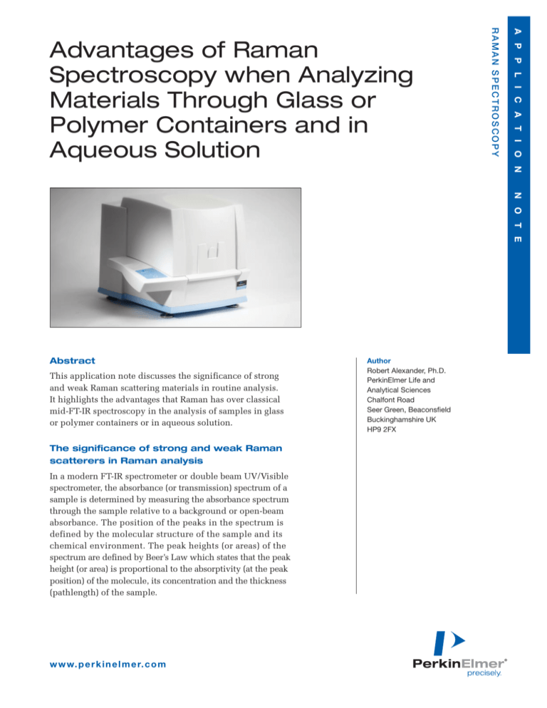
scattering. For instruments such as the RamanStation™
400 which have a relatively weakly focused laser beam,
bulk materials can be measured and analyzed easily
through polymer containers with little or no contribution
from the container. Figure 7 compares the spectrum from
paracetamol taken through a polyethylene bag with the
spectrum from pure paracetamol powder. Only the small
contribution at 2840-2890 cm-1 from the polyethylene
bag is evident in the top spectrum. If required, the contribution from the polymer container can be removed
by automatic spectral subtraction but this is rarely
required for positive material identification.
Figure 8. Analysis of ethanol in thick plastic solvent bottle.
Raman spectroscopy is therefore an extremely good
and easy technique for the analysis of materials in
either glass or polymer containers. These analyses
are particularly important where:
1) speed and convenience of analysis is important such
as in a QC/QA laboratory
2) samples are toxic or have an unpleasant smell
3) samples may be unstable to air or moisture
4) it is important to maintain the safety of the analyst
such as when working with active pharmaceutical
materials
5) the properties and toxicity of the material is unknown
such as at the scene of an accident
6) confiscated forensic materials of unknown composition
or history are being analyzed
7) the integrity of the material is paramount such as in
many forensic analyses
Quantitative Raman analysis in solution
In UV/Visible and IR spectroscopy, the spectral contribution
from the instrument is monitored and removed by using
a reference beam (in a double-beam instrument) or by
subtracting a background spectrum (in an FT instrument).
Figure 7. Paracetamol in polyethylene bag.
Raman analyses can also be made through thicker
polymer containers such as solvent bottles. Figure 9
shows the Raman spectrum of ethanol taken through
a 1.5 mm thick plastic solvent bottle. In this case, there is
4
Since Raman spectroscopy is essentially a single-beam
technique, any contribution or variation due to the instrument must be considered when doing quantitative
analyses. Potential sources of instrumental and sampling
variations are laser and detector stability and de-polarization effects. For this reason, it is vital when doing
quantitative analyses to have as stable an instrument
as possible and to ensure consistency in measurement
parameters (such as laser power, laser focus on sample,
scan sequence, spectral resolution, sample orientation)
and in any post-run processing (such as baseline correction,
noise reduction). This is similar to any quantitative
analysis where any sources of unwanted variation are
kept to a minimum.
It is also more straightforward to do quantitative analyses
on liquids and solutions than on solids since they are
more homogeneous and without the problems associated
with variable particle sizes and shapes.
For these reasons it is more common to measure relative
concentrations of the components in a mixture with
band-ratio methods than with absolute quantitative
analyses. It is also recommended that a standard reference mixture is measured as part of each analysis
regime simply to monitor the stability of the system.
Provided the above considerations are adhered to,
Raman can be a very useful quantitative analysis technique particularly for aqueous samples or samples in
glass or polymer containers which can be difficult to
analyse by IR. This is because relatively strong Raman
scatterers can be measured in the presence of weaker
Raman scatterers. An example of this is the measurement of ethanol in water in a glass container (Figure 9).
Figure 10. Raman spectra of different ethanol concentrations.
Table 1. Peak Heights of Ethanol at Various Concentrations in
Water.
Concentration of
Ethanol in water
Peak Height (880 cm-1
relative to baseline
905-850 cm-1)
0.25%
65.6
1.25%
350.1
5.00%
1423.6
The Raman spectra of several alcoholic beverages were
measured under the same scan conditions (Figure 11)
and the peak heights for the ethanol measured (Table 2).
Once again, these peak heights show a good correlation
with known alcoholic content for these drinks. A more
exact quantitative analysis for these drinks would require separate calibration for each drink type to take
into account variation in their composition. The absolute peak height values in Table 2 do not match with
corresponding values in Table 1 because the scan
conditions were different for the two sets of data.
Figure 9. Spectrum of ethanol showing strong, sharp band at 880 cm-1.
The peak at ~880 cm-1 is an ideal band to be used in a
quantitative analysis. Figure 10 shows the variation in
the height of this peak with varying concentration of
ethanol. There is some variation in baseline position but
this is easily compensated for by measuring the peak
height relative to an adjacent baseline position. These
peak height values are given in Table 1 and illustrate a
good degree of linearity.
Figure 11. Raman spectra of whisky, wine and cider.
w w w. p e r k i n e l m e r. c o m
5
Table 2. Peak Heights of Ethanol in a Variety of Alcoholic
Drinks.
Alcoholic Drinks
Peak Height
Scotch whisky (40%)
87208
White wine (9%)
18087
Cider (6%)
11731
Conclusions
The intensity of Raman spectra not only depends on the
molecular and physical properties of the material but
also on the analysis conditions. Molecules can be classified as strong or weak Raman scatterers depending on
their molecular structure. Experimental parameters such
as laser power and scan times will also influence the
strength of the resultant spectrum. This means that the
analyst must take into account both the molecular structure and the scanning conditions when comparing
Raman spectra from different samples.
Thin samples such as polymer films or coatings are best
measured using a Raman microscope system where the
laser power can be concentrated onto these thin samples
to minimize unwanted contribution from the surrounding
materials.
The fact that glass, polymer films and water have relatively weak Raman spectra when measured using the
RamanStation means that this instrument is ideal for the
analysis of bulk materials in glass or polymer containers,
or in aqueous solution.
Provided adequate experimental procedures are observed,
Raman spectroscopy can be used very effectively for
quantitative analyses in situations where mid-IR is not
appropriate.
PerkinElmer, Inc.
940 Winter Street
Waltham, MA 02451 USA
Phone: (800) 762-4000 or
(+1) 203-925-4602
www.perkinelmer.com
For a complete listing of our global offices, visit www.perkinelmer.com/lasoffices
©2008 PerkinElmer, Inc. All rights reserved. The PerkinElmer logo and design are registered trademarks of PerkinElmer, Inc. RamanStation is a trademark of PerkinElmer, Inc. or its
subsidiaries, in the United States and other countries. All other trademarks not owned by PerkinElmer, Inc. or its subsidiaries that are depicted herein are the property of their respective
owners. PerkinElmer reserves the right to change this document at any time without notice and disclaims liability for editorial, pictorial or typographical errors.
008092_01


