Respiratory System
advertisement

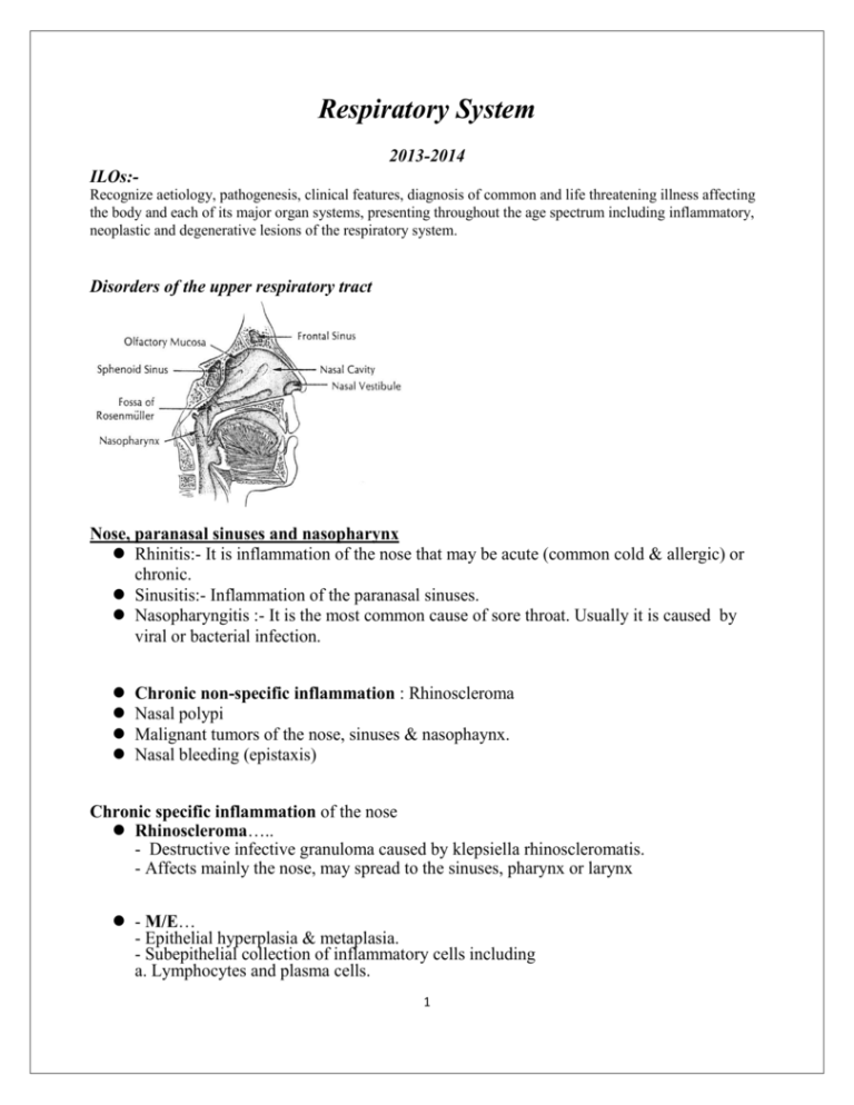
Respiratory System 2013-2014 ILOs:Recognize aetiology, pathogenesis, clinical features, diagnosis of common and life threatening illness affecting the body and each of its major organ systems, presenting throughout the age spectrum including inflammatory, neoplastic and degenerative lesions of the respiratory system. Disorders of the upper respiratory tract Nose, paranasal sinuses and nasopharynx Rhinitis:- It is inflammation of the nose that may be acute (common cold & allergic) or chronic. Sinusitis:- Inflammation of the paranasal sinuses. Nasopharyngitis :- It is the most common cause of sore throat. Usually it is caused by viral or bacterial infection. Chronic non-specific inflammation : Rhinoscleroma Nasal polypi Malignant tumors of the nose, sinuses & nasophaynx. Nasal bleeding (epistaxis) Chronic specific inflammation of the nose Rhinoscleroma….. - Destructive infective granuloma caused by klepsiella rhinoscleromatis. - Affects mainly the nose, may spread to the sinuses, pharynx or larynx - M/E… - Epithelial hyperplasia & metaplasia. - Subepithelial collection of inflammatory cells including a. Lymphocytes and plasma cells. 1 b. Mickulicz cells…hydropic degeneration in pasma cells. c. Russel bodies…hyaline change in plasma cells. - Granulation tissue and fibrosis Complications a. Nasal obstruction b. Epistaxis c. Ulceration and secondary infection. d. Spread of infection Nasal polyps 1- Inflammatory polyps… - Finger-like projections. - Mostly represent swollen parts of inflamed nasal mucosa. - Typically found in patients with recurrent attacks of allergic rhinitis ( have large numbers of eosinophils) or chronic infections of the nasal sinuses. 2- Squamous papilloma… - Benign epithelial tumors. Formed of fibrous core covered by hyperplastic stratified squamous epithelium. 3- Juvenile angiofibroma - Originates from the nasopharynx and projects into the nose. - Benign tumors of young men. May be related to testosterone . - They are composed of thin walled blood vessels within loose fibrous stroma. - They bleed easily and cause epistaxis. 2 Malignant tumors of the nose and Nasopharynx 1- Nasopharyngeal carcinoma 2- Squamous cell carcinoma (Keratinizing and non keratinizing) 3- Adenocarcinoma (5%) of nasal malignant tumors. 4- Plasmacytoma Nasopharyngeal carcinoma - Most common nasopharyngeal tumor, located on the roof of the pharynx. - Males affected more than females. - Associated with Epstein Barr Virus infection especially in China - Histologically it is composed of undifferentiated squamous cells typically intermixed with non-neoplastic lymphocytes (lymphoepithelioma). - Early nodal metastases. - Treatment by surgery and irradiation give good results (5 years survival rate 60%). Nasal bleeding (epistaxis) - Causes 1- Local….. a. Trauma b. Foreign body c. Weak Little’s area d. Inflammation e. Tumors (benign or malignant) 2- General a. Hypertension b. Generalized venous congestion c. Blood disease d. Anticoagulant therapy e. Vitamin deficiency (K and C) 3 Paranasal sinuses Sinusitis - It is inflammation of the para nasal sinuses, caused by extension from nasal cavity or dental infections. - It results in obstructed drainage from the sinuses with accumulation of mucous secretion and secondary bacterial infection. Larynx Laryngitis Inflammation of the larynx may be caused by infections (virus or bacteria), irritation or over use of the voice. - Inflammation and edema of the vocal cords cause hoarseness of voice. Tumors of the larynx 1- Singer's nodule… - Small benign laryngeal polyp. - Usually associated with chronic irritation of the vocal cords as excessive use of voice and heavy smoking. - They are located on the true vocal cords and present by hoarseness of voice. 2- Laryngeal papilloma.. - true benign neoplasms of the larynx. a. In adults, the neoplasm is usually single, do nor recur after removal but may undergo 4 malignant changes. b. In children (Juvenile papillomatosis) they are multiple, caused by human papilloma virus, recur after removal but do not undergo malignant changes 3- Squamous cell carcinoma….. most common malignant tumor of the larynx. - Predisposing factors include - Smoking - Asbestos and chromium. - Adult laryngeal papilloma. - Leukoplakia - Pathology May be a. Verrucal carcinoma - Uncommon, low grade papillary squamous cell carcinoma. - It is superficially invasive . – Relatively of good prognosis Verrucal carcinoma b. Classic invasive squamous cell carcinoma - Usually affects males above 40 years (Male : female 7:1). - Related to smoking and alcoholism. According to the site they may be - Glottic carcinoma… arise on the true cord so the patient presents early by hoarseness of voice. - It is the most common (60-70%) and has the best prognosis. Glottic carcinoma 5 - Supraglottic and subglottic….less common, present late, have poorer prognosis. - Treatment by surgery and irradiation give good results (5 years survival rate 60%). Supra glottic carcinoma Tonsils The mucosa associated lymphoid tissue around the pharynx (Waleyer's ring) comprises the palatine tonsils (tonsils), the nasopharyngeal tonsils (adenoids), and the lymphoid tissue in the submucosa of the posterior third of the tongue (lingual tonsil). - They are prominent in children and tend to be fibrosed with advanced age. - These lymphoid aggregates react to inflammation or infection by reactive hyperplasia. A. Adenoids - Inflammatory hyperplasia of the nasopharyngeal lymphoid tissue,affecting mostly children. - Hyperplastic enlargement leads to nasal obstruction and mouth breathing. - If the case is neglected, it leads to adenoid facies ( narrow nasal openings, open mouth,short upper lip and absent naso-labial folds). - Secondry bacterial infection with spread to the middle ear leads to otitis media and also to lower respiratory tract infection. Adenoid face B. Palatine tonsils (tonsils) - Acute inflammation a. Diphtheria b. Acute tonsillitis 6 a.Diphtheria - Due to infection by Corynebacterium diphtheriae. - It is now very rare due to vaccination. - It is a peudomembranous type of inflammation. - It can lead to obstructive asphyxia in children and for the production of bacterial exotoxin that affects the heart (toxic myocarditis) and nervous system. b. Acute tonsillitis - Usually occurs as a component of acute bacterial pharyngitis and caused mostly by βhemolytic streptococci. - Inflammation is bilateral and passes into a. Catarrhal inflammation b. Acute follicular tonsillitis …acute suppurative inflammation of the lymphoid follicles with pus collecting into the tonsillar crypts then spread to cover the surface. Acute follicular tonsillitis (Complications) 1- Peritonsillar abscess (quinsy). 2- Rarely spread of infection causing neck cellulites (Ludwig's angina). 3- Retropharyngeal abscess. 4- Otitis media. 5- Blood spraed (septicemia or pyemia) 6- Allergic diseases ( Rheumatic fever or post-streptococcal glomerulonephritis). 7- Chronicity. Lower Respiratory System - Anatomically - The right lung has three lobes and the left lung is divided into two lobes ( the middle lobe equivalent is called the lingual). Both main bronchi arise from the trachea and then branch to progressively smaller airways. Progressive branching of the bronchi form bronchioles, which in contrast to bronchi lack cartilage and submucosal glands in their walls. Bronchioles further branch giving rise to terminal bronchioles. A cluster of 3-5 terminal bronchioles and their appended acini form the pulmonary lobule. 7 Pulmonary acinus The structural unit of the lung distal to the terminal bronchiole is called the acinus. The acinus is formed of respiratory bronchioles, alveolar ducts and alveolar sacs. Histologically - Most of the respiratory tree is lined with pseudostratified, tall columnar, ciliated epithelial cells with mucus secreting goblet cells admixed with cartilaginous airways. - Bronchial mucosa also contains neuro-endocrine cells. - Numerous submucosal mucus secreting glands are dispersed throughout the walls of trachea and bronchi (but not bronchioles). Atelectasis and Collapse I- Atelectasis = (atelectasis neonatorum) Definition: it is incomplete or non-expantion of lung alveoli in newborn. Causes: A- Primary atelectasis: of unknown cause. B- Secondary atelecasis due to. 1- Prematunity ( surfactant). 2- In full term fetus, it is due to: a- Cerebral causes: Cerbral bith injuny e.g. - Traumatic intracranial haemorrhage compresses respiratory center. - Anaethesia to the mother. - Coiling of umblical cord around neck of foetus. b- Lung causes include: -Congenital syphilis. - Congenital bronchial obstruction. - Amniotic fluid aspiration. 8 ii- Collapse Definition: It is deflation of fully areated alveoli (i.e after complete inflation). It usually occurs in adult. Causes and types: A- Obstructive (absorptive) collapse B-Compression (pressure) collapse C- Contraction collapse A- Obstruction atelectasis Obstruction of an airway prevents air from reaching distal airways and the air already present gradually becomes resorbed leading to collapse. Causes of obstruction include - Inside the bronchus …. Mucous , blood clot, foreign body, tumor. - Outside the bronchus ..enlarged lymph node, aneurysm. B-Compression (pressure) collapse: Due to external pressure on the lung which pushes the air outside the alveoli. It is caused by: Pleural causes: air (Peumothorax) pus (Pyothorax) blood (haemothorax) lymph (chylothorax) serous fluid (hydrothorax). Extra pleural causes: Elevated diaphragm by ascitis or subdiaphragmatic abscess. Mediastinal tumour 3- Dilated heart. Spinal deformity 9 C- Contraction collapse Occurs when local or generalized fibrotic changes impair lung expansion. It is an irreversible type of atelecasis Pathology of both atelectasis and collapse. N/E: (1) Massive (complete or total). - Affects the whole lung. The lung is small and bluish (due to reduced HB). The pleura are wrinkled. (2) Partial or incomplete: - Affects part of the lung. e.g lobular or lobar. The affected area is depressed below the surrounding surfaces. It appears triangular in shape with the base at the pleura and apex centrally. It is bluish in colour, rubbery in consistency and does not cripitate. M/E: The alveolar walls are approximated together. The alveolar spaces are narrowed to slit-like openings (may contain oedema fluid). The inter-alveolar septa are thickened and their capillaries are dilated and congested due to loss of compressive force of air. There is an apparent increase in the number of bronchioles. + in atelectasis the alveolar spaces are lined by cuboidal epithelium (foetal lung). Complications: 1- lung infection. 2- Respiratory distress. 3- Pulmonary fibrosis in prolonged cases. 4- Pulmonary hypertension (due to fibrosis), and cor pulmonale. Lung diseases Include:1- Obstructive and Restrictive Diseases. 2- Pulmonary Vascular Diseases.(congestion, infarction,embolism….etc) 3- Pulmonary Infections. 4- Pulmonary Neoplasia. 5- Diseases of the pleura 10 Obstructive and Restrictive lung diseases Diffuse lung diseases can be classified into two groups 1- Obstructive (airway) diseases characterized by limitation of pulmonary airflow. 2- Restrictive diseases characterized by reduced lung expansion with decrease of the total lung capacity. Obstructive and Restrictive lung diseases Obstructive Restrictive Normal or increased Reduced capacity &FVC FEV1 Decreased Normal or reduced FEV1/FVC Decreased Near normal Examples 1- Asthma 2- Emphysema 3- Chronic bronchitis and bronchiolitis. 4- Bronchiectasis - Acute and chronic interstitial lung disease 1- ARDS 2- Pneumoconiosis 3- Sarcoidosis. 4- Idiopathic pulmonary Total lung 5- Cystic fibrosis fibrosis Chronic Obstructive Pulmonary Diseases Definition: A group of pulmonary diseases characterised by increased resistance to air flow due to partial or complete obstruction at any level. Types: 1) Chronic bronchitis. 2) Bronchial asthma. 3) Emphysema. 1- Chronic Bronchitis Definition: - It is persistent productive cough (cough of sputum) for at least 3 consecutive months in at least 2 consecutive years. Causes: Due to chronic irritation of the bronchial mucosa by: 1- Cigarette smoking. 2- Atmospheric pollution 3- Chronic inflammation of upper respiratory tract, tonsils or mouth. Pathology: Occurs in middle-aged men. 11 Grossly - Mucosal lining of large airways show edema, hyperemia and is covered by mucus or muco-purulent secretion. - Small bronchi and bronchioles may show the same picture. Microscopically 1- Enlargement of the mucus-secreting glands with increased Reid index ( ratio between mucous gland layer to the wall). 2- Increased number of goblet cells in the lining epithelium with loss of ciliated cells. 3- Squamous metaplasia of the epithelium which may be typical or dysplastic 4- Infiltration of the wall by chronic inflammatory cells . 5- In chronic bronchiolitis there is goblet cell metaplasia of small airways with inflammation and fibrosis of their walls, and smooth muscle hyperplasia….all leading to small airway obstruction. 12 - Inflammation and mucus secretion result in airway obstruction. Complications: 1- Centrilobular emphysema. 2- Bronchopneumonia. 3- Bronchogenic carcinoma. 4- Chronic hypoxaemia resulting in persistent pulmonary vasoconstriction, pulmonary hypertension and corpulmonale. 5- Cardiac failure. 2- Bronchial Asthma Definition: A common disease characterised by recurrent attacks of widespread broncho-constriction. in response to various stimuli. Aetiology and Types: (1) Extrinsic (immunological, atopic, allergic) asthma: - 10% of cases. - Affects children and young adults. - A positive family history of asthma or atopic disease is common. - Attacks are provoked by type I hypersensitivity reaction to inhaled allergens such as house dust, animal dandruffs, plant pollens, fungi, ….etc (2) Intrinsic (non-immunological, non-atopic, non allergic) asthma: - About 90 % of cases. - It occurs at any age and mainly in late adults. - No history of atopic disease. - It is triggered by hyperirritability of bronchial tree by infection , exposure to cold, physical exercise, anxiety, emotions, asprin and inhaled irritants... etc. 13 Morphology Grossly - Obstruction of bronchi and bronchioles by thick tenacious mucus plugs. - Lung tissue shows areas of collapse alternating with areas of hyperinflation. Microscopically:x From inside outwards 1- The lumen of bronchi and bronchioles contains excess mucous with scattered esinophils. 2- The epithelium shows goblet cell hyperplasia with thickening of the basement membrane. 3- The subepithelial tissue contains excess mixed inflammatory infiltrate (lymphocytes, macrophages, neutrophils, esinophils asnd mast cells) 4- Smooth muscle hypertrophy 5- Mucous gland hyperplasia 6- Sputum examination shows Charcot-Leyden crystals and Curschmann's spiral Curschmann's spiral Charcot-Leyden crystals 14 Fate and Complications: l - Chronic bronchitis. 2- Emphysema, pulmonary hypertension and Rt. sided heart failure. 3- Status asthmaticus: persistent attacks for days or weeks. It is fatal due to respiratory failure. Emphysema Definition Emphysema is a lung condition characterized by abnormal permanent enlargement of the airspaces distal to the terminal bronchiole, accompanied by destruction of their walls without obvious fibrosis. Incidence - A common disease. - Males > females. - More in smokers (centriacinar type) especially heavy smokers. - Becomes disabling by the fifth decade of life. 15 Types of emphysema The forms of emphysema are defined by their anatomic nature within the acinus. There are many types of emphysema 1- Centriacinar 2- Panacinar 3- Distal acinar (paraseptal) 4- Irregular 1- Centriacinar (centrilobular) emphysema - The central or proximal parts of the acini, formed by respiratory bronchioles, are affected, where as the distal alveoli are spared. - It is usually more severe in the upper lobes especially the apical segments. - It tends to occur in heavy smokers and the walls of the emphysematous spaces often contain abundant black pigment. Pathogenesis The current opinion favors emphysema arising as a consequence of two critical imbalances 1- Protease-antiprotease imbalance 2- Oxidant-antioxidant imbalance. 16 2- Panacinar (panlobular) emphysema - Is characterized by uniform enlargement of the acini from the level of the respiratory bronchioles to the terminal blind alveoli. - It affects the lower lobes and anterior margins of the lungs more severely - It is associated with alpha1-antitrypsin deficiency. 3- Distal acinar (paraseptal) - The proximal part of the acinus is normal and the distal acinar portion is mainly affected. - It is more severe in the upper half of the lungs adjacent to the pleura. - A characteristic feature is the presence of multiple, enlarged airspaces ranging in diameter between 0.5 to more than 2.0 cm, sometimes forming cyst like structures called bullae that may rupture into the pleura leading to pneumothorax. Morphology Grossly - In Centriacinar emphysema the lungs are less voluminous, more pink with black pigmentation (due to smoking). The upper parts of the lungs tend to be more affected and 17 - In severe cases, of any type of emphysema especially the distal acinar or panacinar types, emphysematous bullae may be visible. - In panacinar emphysema the lungs are voluminous, pale and often obscure the heart. The lower lobes tend to be more affected. Panacinar emphysema l) Barrel shaped chest: The chest wall takes a fixed exaggerated inspiration position: a- The anteroposterior diameter increases to equal the transverse. The sternum is pushed forward and moderate kyphosis occurs. b- The ribs, costal cartilages and the intercostal spaces are horizontal. c- The subcostal angle is wide. Microscopically - Thinning and destruction of alveolar walls .Adjacent alveoli become confluent creating large air spaces. - Diminished number of alveolar capillaries. - Evidence of bronchitis and bronchiolitis 18 Complications 1- Pulmonary hypertension due to a. Hypoxia leading to pulmonary vascular spasm. b. Decreased capillaries due to alveolar wall destruction - Pulmonary hypertension leads to right sided heart failure (cor pulmonal). 2- Pulmonary bacterial infections. 3- Pulmonary failure, respiratory acidosis and cyanosis. 4 - Pneumothorax. Conditions related to emphysema 1- Compensatory emphysema 2- Senile emphysema 3- Obstructive over inflation 4- Interstitial (mediastinal) emphysema Interstitial Emphysema It is the escape of alveolar air into the interstitial tissue of the lung. Causes: 1) Alveolar tear in pulmonary emphysema due to sudden increase of intra alveolar pressure e.g. severe coughing, lifting a heavy weight, straining. 2) Physical injury due to instrumentation of airways, fractured rib or chest wound penetrating lung Fate: 1) Blood clots seals lung tears and the escaped air is slowly absorbed into the blood. 2) If the amount of air is extensive it may: a- Compress the blood flow. b- Produce air embolism. c- Produce pneumothorax. 19 Bronchiectasis Definition: Permanent (irreversible) dilatation of medium sized bronchi and bronchioles caused by chronic suppurative inflammation in their walls and surrounding lung tissue. Incidence: It is frequent before the age of 20 years in males more than females. However, it can occur in adults. Causes a. Bronchial obstruction - Localized (tumors; foreign bodies), - Diffuse (atopic asthma; chronic bronchitis) b. Congenital or hereditary conditions (Few cases e.g. cystic fibrosis) c. Necrotizing or chronic suppurative pneumonias. - Especially with virulent organisms (staph aureus, klepsiella and tuberculosis Pathogenesis Two factors each of them leads to the other, any of them may come first 1- Airway obstruction leading to a. Retention of secretions….Favors secondary bacterial infection and increases intraluminal pressure. b. Atelectasis.. pulling on the bronchi and bronchioles from the outside. 20 2- Infection leading to a. Increased secretions leading to further obstruction. b. Destruction of walls followed by peri-bronchial fibrosis with loss of elastic and muscular tissue …leading to weakening and dilatation of the walls. Grossly - Affection is usually bilateral and basal with more severe affection of distal bronchi and bronchioles. - Airways are dilated and can be traced up to the pleura. This dilatation may be saccular, fusiform or cylindrical. - If obstruction is due to tumor or foreign body, bronchiectasis is sharply localized to a single segment. M/E - The affected bronchi and bronchioles show:a. The lumen contains exudates and pus cells b. Shedding and ulceration of the epithelium. c. Acute and chronic inflammatory cells in the wall. 21 d. Destruction and fibrosis of the wall and peri-bronchial tissue with abnormal dilatation and scarring of bronchi and bronchioles. e. Collapse of surrounding lung tissue. Clinical course -The clinical picture is chronic cough with mucopurulent fetid sputum production. - Sometimes hemoptysis is evident. – Clubbing of the fingers may develop. - In late, advanced cases, obstructive manifestations are evident with hypoxemia and cyanosis. Complications 1- Lung abscess, and lung gangrene 2- Pulmonary hypertension and cor pulmonale. 3- Spread of infection leading to pyemia and brain abscesses or septicemia. 4- Reactive systemic amyloidosis, Restrictive lung diseases A group of lung diseases characterized by reduced expansion of the lung and reduction of the total lung capacity. - The FVC is reduced and the FEV1 is normal or reduced and so the FEV1/FVC is normal. Causes 1- Abnormalities of the chest wall that restrict lung expansion - Bony abnormalities…kyphoscoliosis - Neuro-muscular diseases ..poliomyelitis - Severe obesity. 2- Pulmonary causes (Acute and chronic Interstitial lung diseases) 1- Acute (Adult) and neonatal respiratory distress syndromes. 2- Pneumoconiosis 3- Hypersensitivity pneumonitis. 4- Idiopathic pulmonary fibrosis 5- Sarcoidosis 6- Diffuse alveolar hemorrhage syndromes 7- Pulmonary angiitis and granulomatosis 8- Lung affection in collagen diseases 22 -Pneumoconiosis - It is a group of environmental or occupational lung diseases cause by inhalation of inorganic dust particles. They include A. Anthracosis is caused by inhalation of carbon dust, it is endemic in urban areas and causes no harm. - It is characterized by carbon - carrying macrophages resulting in irregular black areas seen on gross or microscopic examination of the lungs. B. Coal workers’ pneumoconiosis is produced by inhalation of coal dust which contains both carbon and silica. - It may be complicated by bronchiectasis, pulmonary hypertension , respiratory failure or right –sided heart failure. C. Silicosis is a chronic occupational lung disease caused by exposure to free silica dust. It is seen in miners, glass manufactures and stone cutters. - The disease is mediated by ingestion of silica particles by alveolar macrophages, damage of these macrophages initiates an inflammatory reaction followed by fibrosis - The lungs show silicotic nodules that obstruct the airways and blood vessels. - Silicosis is associated with increased susceptibility to tuberculosis. D. Asbestosis is caused by inhalation of asbestos fibers. - The disease is initiated by uptake of asbestos fibers by alveolar macrophages. A fibroblastic response occurs due to release of fibroblast-stimulating growth factors by macrophages leading to diffuse interstitial fibrosis mainly in the lower lobes. - The lung lesion is characterized by ferruginous bodies formed of asbestos fibers coated with iron and protein. 23 - The pleura shows hyalinized fibrocalcific plaques on the parietal pleura. - Asbestos results in marked predisposition to bronchogenic carcinoma and malignant mesothelioma of the pleura and peritoneum. -Cigarette smoking further increases the risk of bronchogenic carcinoma. - Hypersensitivity pneumonitis - It is a group of immunologically mediated conditions caused by intense, often prolonged exposure to organic dusts. - It is important to recognize these diseases early because progression to chronic fibrotic lung diseases can be prevented by removal of the environmental agent. - This group includes - Farmer’s lung.. inhalation of spores of actinomycetes bacterial when hay or corn is gathered wet. - Byssinosis… Inhalation of cotton dust - Bagassosis… Inhalation of bagasse residue after sugar juice extraction - Pigeon breeder’s lung - Humidifier or air conditioner lung. inhalation of actinomycetes Lung infections Pneumonia Means inflammation of the lung tissue. It may be acute or chronic. - It occurs when the invading organism overcomes the innate defense mechanisms of the respiratory tract. - It is characterized clinically by fever, chills, productive cough, blood tinged (rusty) sputum, pleuritic pain, may be hypoxia and cyanosis. - If pneumonia is of bacterial origin, it is associated with neutrophilia. 24 Pneumonia Inflammation of lung tissue. Classification 1) Bacteria1: - Lobar pneumonia. - Bronchopneumonia. 2) Primary Atypical Peneumonitis: It is an acute interstitial inflammation confined to alveolar septa without intra-alveolar exudate. Causes: 1- Viruses: as influenza, parainfluenza, measles chicken pox and small pox. 2- Mycoplasma pneumonia. 3- Undefined agent. 3) Loeffler's Pnueumonia: It is eosinophilic pneumonia with eosinophilia. It is due to parasitic infestations e.g. ascaris, ankylostoma and Bilharziasis (verminous pneumonia). 4) Granuloma: as T.B., sarcoidosis, leprosy, actinomycosis, moniliasis. 5) Lipid Pneumonia: It is pneumonic consolidation due to lipid accumulation, Source of lipid: Endogenous from degenerated lesions as cancer or T. B. 25 Exogenous, usually due to oily nose drops. 6) Irradiation Pneumonia: Ionising radiation producese diffuse pulmonary fibrosis. Bacterial Pneumonias - They are bacterial in origin and may follow viral upper respiratory tract infection. - In most cases it is caused by Steptococcus pneumonae infection. - It can produce either lobar pneumonia or bronchopneumonia. A. Lobar pneumonia - It is an acute bacterial pneumonia caused in 90% of cases by infection by S.pneumonae.( gram-positive diplococcus) - There is contiguous affection of part or whole lung lobe homogenously. - It is a sero- fibrinous or sero-purulent type of inflammation. - It usually affects middle aged. Morphology - Part of the lung lobe or whole lobe is affected. - Lower lobes or right middle lobe are more commonly affected . - In the pre-antibiotic era, the disease passes into 4 stages:1- Congestion (1-2 days): - Blood vessels dilate and leak fluid in response to injury. - Affected lobe becomes heavy, red and boggy. - M/E… - Congested alveolar capillaries. - Alveolar spaces contain edema fluid, bacteria and few neutrophils 26 2. Red hepatization: (2- 4 days): - The inflammation progresses, and the affected lobe has a consolidated (airless) liver like consistency. - The overlying pleura shows serofibrinous inflammation. - M/E…. Congested , damaged interalveolar capillaries. - Alveolar spaces contain fibrin meshwork, RBCs, many polymorphs and bacteria - The alveolar exudate becomes "rusty sputum". - The overlying pleura shows serofibrinous or fibrino-suppurative inflammation 3. Grey hepatization (4-8 days) : - Fibrin dominates the picture, while polymorphs and red cells break down ("grey" because hemorrhage is no longer taking place and the red cells have lysed) - The lung becomes dry, firm and grey. 27 - M/E…Less congestion, less edema and rbcs. - Alveoli contain fibrin network which starts to shrink, entangle bacteria , polymorphs 4-Resolution (8 - 21 days): Plasmin clears out the fibrin, and the lung returns to normal in non complicated cases . - Enzymatically digested exudates are either phagocytosed by alveolar macrophages or expectorated leaving the lung structure intact (resolution). - The pleural reaction may undergo either resolution or organization by fibrous tissue leading to thickening and adhesions B. Bronchopneumonia - Caused by a wide variety of organisms. - The process of inflammation spreads from the bronchioles to the adjacent alveoli. - There is patchy distribution in one or more lobes mostly the affection is bilateral and basal. - It usually affects the extremes of age (very young and very old). - Patchy consolidation in one or more lobes preferably bilateral and basal. - Affected patches are 3-4 cm. in diameter, slightly elevated, red, yellow or grey in color surrounded by edema and hyperemia. - Other lung tissue may show hyperinflation or may be normal. - Overlying pleura is less severely affected than in lobar pneumonia - M/E…Focal suppurative inflammation in bronchi, bronchioles and surrounding alveolar spaces. - No distinct stages of inflammation as in lobar pneumonia. 28 - Diagnosis depends upon sputum culture and sensitivity and choosing the proper antibiotic therapy. Prognosis and Complication - With appropriate antibiotic therapy , complete restitution of lung is the rule in both pneumococcal lobar pneumonia and bronchopneumonia. - Occasional cases may be complicated (especially with type 3 pneumococci) with:1- Abscess formation due to extensive tissue destruction and necrosis.(post-pneumonic lung abscess) 2- Affection of the pleura a. The pleural surfaces overlying the infection are almost always involved, accounting for the pain of lobar pneumonia. There will be fibrinous adhesions, which may resolve or turn into scars. b. Suppurative inflammation of the pleura with accumulation of pus inside resulting in empyema Organized pneumonia 3- Non- resolution of the intra-alveolar exudates with organization resulting in lung fibrosis 4- Spread of infection to other parts of the body due to bacterial dissemination leading to meningitis, arthritis, and infective endocarditis. N.B: Today, uncomplicated lobar pneumonias are easily treated with antibiotics once the etiologic agent is identified. Lung abscess - It is a localized suppurative inflammation characterized by necrosis of the lung tissue. - The causative micro-organisms (aerobic and anaerobic streptococci, S.aureus and gram negative organisms) are introduced into the lung by several mechanisms Types and causes of lung abscess ① Aspiration of infected material from the upper respiratory tract, oral cavity or aspiration of gastric contents ( in cases where the cough reflex is inhibited in alcoholics, coma , anesthesia, oral sepsis…) ② Pre-existing lung infection ..pneumonias, bronchiectasis. ③ Septic emboli (pyemic abscess) 29 ④ Obstruction ..most commonly by a tumor. ⑤ Direct penetrating injury or spread from an adjacent structure (rib). Grossly - Abscess may be single (aspiration, obstruction, direct spread) or multiple (post pneumonic or pyemic). - Size varies from few mms to up to 6 cm in diameter. - Site depends upon the cause (aspiration cause right , single abscess- postpneumonic are scattered small multiple bilateral basal- embolic are small, bilateral scattered anywhere within the lungs) M/E… as any abscess with lung tissue destruction and surrounding fibrosis. Complications - Empyema - Brocho-pleural fistula - Pyothorax and pyoneumothorax. - Spread of infection to extra pulmonary sites either directly or by hematogenous spread . - Lung fibrosis leads to pulmonary hypertension, right sided heart failure. - Chronic abscesses lead to clubbing of the fingers and secondary amyloidosis. 30 Tumors of the lung A- Benign Tumors:- Only a small minority of total lung tumors. - Most of them are hamartomas. - They present as small (2-6 cm) white firm nodules. - Formed of cartilage with clefts lined by respiratory epithelium with areas of fatty, fibrous tissue and blood vessels. - Appear in chest X ray as coin lesion (important only in D.D with malignant lesions). Low Grade Malignant Tumors - This group includes - Carcinoid tumor ( typical and atypical) - Adenoid cystic carcinoma - Mucoepidermoid carcinoma. Carcinoid tumor - A tumor of neuro-endocrine origin may be part of MEN-1 syndrome. ◈ Incidence - About 5% of all pulmonary neoplasms. - Age … younger than 40 years. - Sex ….equal in both - Not related to smoking or other environmental factors. Grossly - Typical carcinoids are located in the large bronchi, - Small (3-4 cm) lesion. May be intraluminal (intrabronchial) nodule covered by intact mucosa, or it may present as a penetrating mucosal plaque to the peribronchial tissue with a pushing border. 31 M/E: - Nests of small uniform cells with regular rounded nuclei , absent or few mitotic figures separated by scanty stroma. - Tumor cells are positive for neurone-specific enolase and other neuro-endocrine markers. - Few tumors show high mitotic counts, more atypia, more nodal metastases and a worse overall prognosis ( Atypical carcinoid) Clinical behavior - Patients present with cough, hemoptysis and bronchial obstruction. - Radiologically the tumor appears as coin lesion. - Rare patients with bronchial carcinoid present with carcinoid syndrome (flushing, diarrhea bronchial spasm ). - A slowly growing, mostly surgically curable tumor. - About 10-15% of patients have hilar L.N metastasis by the time of diagnosis (with no change in the prognosis). Rarely the tumor sends distant metastasis. - 5-10 years survival rate is 50-95%. C- Malignant Tumors Primary - Bronchogenic carcinoma (90-95% of primary lung tumors). - Others…mesenchymal tumors, lymphomas…. Secondary (metastatic) - More common than primary lung tumors. - Reach the lung either by a. Direct spread…e.g oesophageal tumors b. Lymphatic spread…breast tumors, abdominal malignancy c. Hematogenous spread…liver, genito -urinary,malignant melanoma - Morphology…They are multiple, discrete more to the periphery of the lung. - Metastases from the genito -urinary tract give large spherical metastases (Cannon- ball appearance in X ray). Bronchogenic carcinoma ◈ Epidemiology - Commonest (90-95%) primary malignant pulmonary tumor. - Second most frequent malignancy in western countries. - First leading cause of cancer death in industrialized countries. - Male : Female 2:1 now female incidence is increasing due to increased female smoking habits. - Age…above 50 years. Aetiology and Pathogenesis 32 ①Tobacco smoking … 80% of lung cancers occur in smokers. - Increased incidence with increased number of cigarettes smoked/ day, increased years of smoking . ②Industrial hazards…Radiations, asbestos, nickel, chromate, miners, newspaper workers. ③Air pollution. ④Hereditary (genetic) factor…. Abnormalities of P-450 genetic control. ⑤Molecular background ⓐLoss of tumor suppressor genes - Deletion of 3p……in all types of bronchogenic carcinoma - Mutations of P53 and Rb genes……more with SCLC - Mutation of 16p/CDK more in NSCLC. ⓑOncogene abnormalities - K-ras mutations ….in adenocarcinoma - MYC overexpression…..in both SCLC and NSCLC Histological subtypes of bronchogenic carcinoma 4 Main types 1- Squamous Cell Carcinoma 2- Adenocarcinoma 3- Large Cell Undifferentiated 4- Small Cell Undifferentiated 1 For therapeutic purposes, bronchogenic carcinoma is subclassified into Small Cell Lung Cancer (SCLC) which is not amenable to surgery and Non Small Cell Lung Cancer (NSCLC) in which surgical intervention may be considered Squamous cell carcinoma Central, large bronchus. - Tendency for cavitation ...keratin, squamous pearls, "bridges” Well, moderately or poorly differentiated. 33 2 Adenocarcinoma Small, peripheral tumors related to previous scars. Least relation to smoking. Well, moderate or poorly differentiated Glands, acini &papillae separated by desmoplastic stroma. Bronchiolo-alveolar carcinoma subtype. It is formed of mucin secreting cells. These malignant cells grow along the alveolar septal framework 3- Small Cell Undifferentiated “Small blue cells" (i.e., very little cytoplasm ); EM: "neurosecretory" (dense-core, APUD) granules . Wide areas of necrosis 4- Large Cell Undifferentiated... It may represent poorly differentiated squamous cell or adenocarcinoma 34 Effects of bronchogenic carcinoma • 1- Bronchial obstruction with hyperinflation, atelectasis, and obstructive pneumonia behind a protruding mass. 2- Hoarseness of voice due to Recurrent laryngeal nerve involvement. 3- Pleural and pericardial invasion : resulting in hemorrhagic pleural and pericardial effusion. Spread A. Direct spread - To the pleura and surrounding structures B. Lymphatic spread - To the hilar, mediastinal, scalene and supra-clavicular groups of L.N. C. Hematogenous spread - Bronchogenic carcinoma often involves the brain, bones, liver, adrenals kidneys, heart, pleura, and skin; no organ is immune. Course and prognosis - Most lung cancers are silent until they've become inoperable. Those lucky patients whose cancers are of operable subtype and are detected very early have a reasonably good prognosis . - The onset is gradual. Patients present with cough, chest pain, shortness of breath, and/or (especially) weight loss. The disease is most often unresectable when the patient comes to the physician. - Hoarsness of voice , chest pain, SVC syndrome, pleural or pericardial effusion denote advanced disease. - 3- 10% of patients present with para-neoplastic syndrome which include ⓐEctopic and inappropriate hormone production -ACTH & ADH….. by mall cell carcinoma. - Parathormone….by squamous cell carcinoma. ⓑAcanthosis negricans ⓒClubbing of the fingers. ⓓHematological … migratory thrombophlebitis, and DIC….more with adenocarcinoma - Overall 5 years survival rate is 10% 35 Pleural diseases Pleural Effusion - It- is accumulation of fluid into the pleural space Pathogenesis may be - Increased hydrostatic pressure…..heart failure - Increased vascular permeability……pneumonias - Decreased plasma osmotic pressure…..nephrotic syndrome - Lymphatic obstruction…..malignant tumors. - Increased pleural negative pressure….atelectasis. Causes may be inflammatory or non inflammatory Inflammatory a. Serofibrinous….. Pneumonnias ,uremia, collagen diseases, irradiation. b. Suppurative (empyema)….bacterial or mycotic infections. c. Tuberculous…..exudate contains excess lymphocytes, (> 70% and no mesothelial cells ). Non inflammatory a. Hydrothorax….clear transudate, mostly caused by heart failure, Meig's syndrome, renal or hepatic failure. b. Hemothorax….blood into the plural cavity - May be traumatic, rupture aortic aneurysm or blood disease c. Chylothorax…..accumulation of lymph due to lymphatic obstruction mostly by mediastinal malignancy 36 Pneumothorax - It is accumulation of air into the pleural space, it may be A. Spontaneous pneumothorax… complicating lung diseases as - Emphysema - Asthma - Tuberculosis - Abscess B. Traumatic pneumothorax - Penetrating injury - Fracture rib - Rupture oesophagus - Pneumothorax caused lung compression and collapse with marked respiratory distress. Tumors of the pleura 1- Metastatic tumors - More common than primary pleural tumor. - Primary is usually in the lung or breast. 2- Mesothelioma - Rare rapidly fatal tumors. - Relation to exposure to asbestos. - Consist of proliferating mesothelial cells that may be spindle shaped (sarcomatous), cuboidal and line cleft (carcinomatous) or biphasic (mixed) pattern. 37


