Motor Delays: Early Identification And Evaluation
advertisement
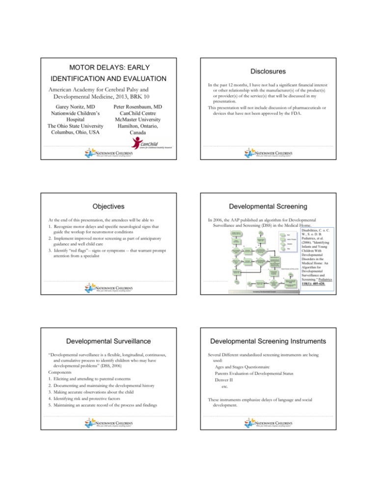
MOTOR DELAYS: EARLY Disclosures IDENTIFICATION AND EVALUATION American Academy for Cerebral Palsy and Developmental Medicine, 2013, BRK 10 Garey Noritz, MD Nationwide Children’s Hospital The Ohio State University Columbus, Ohio, USA Peter Rosenbaum, MD CanChild Centre McMaster University Hamilton, Ontario, Canada In the past 12 months, I have not had a significant financial interest or other relationship with the manufacturer(s) of the product(s) or provider(s) of the service(s) that will be discussed in my presentation. This presentation will not include discussion of pharmaceuticals or devices that have not been approved by the FDA. ………………..…………………………………………………………………………………………………………………………………….. ………………..…………………………………………………………………………………………………………………………………….. Objectives Developmental Screening At the end of this presentation, the attendees will be able to 1. Recognize motor delays and specific neurological signs that guide the workup for neuromotor conditions 2. Implement improved motor screening as part of anticipatory guidance and well child care 3. Identify “red flags”-- signs or symptoms -- that warrant prompt attention from a specialist ………………..…………………………………………………………………………………………………………………………………….. Developmental Surveillance “Developmental surveillance is a flexible, longitudinal, continuous, and cumulative process to identify children who may have developmental problems” (DSS, 2006) Components 1. Eliciting and attending to parental concerns 2. Documenting and maintaining the developmental history 3. Making accurate observations about the child 4. Identifying risk and protective factors 5. Maintaining an accurate record of the process and findings ………………..…………………………………………………………………………………………………………………………………….. In 2006, the AAP published an algorithm for Developmental Surveillance and Screening (DSS) in the Medical Home. Disabilities, C. o. C. W., S. o. D. B. Pediatrics, et al. (2006). "Identifying Infants and Young Children With Developmental Disorders in the Medical Home: An Algorithm for Developmental Surveillance and Screening." Pediatrics ………………..…………………………………………………………………………………………………………………………………….. 118(1): 405-420. Developmental Screening Instruments Several Different standardized screening instruments are being used: Ages and Stages Questionnaire Parents Evaluation of Developmental Status Denver II etc. These instruments emphasize delays of language and social development. ………………..…………………………………………………………………………………………………………………………………….. Why is early diagnosis important? Mean Age First Signs or Symptoms Noted 2.5 years First reported to PCP 3.6 years First Creatine Kinase Sent 4.7 years Definitive Diagnosis of Duchenne 4.9 years 156 boys without a family history of Duchenne (2009) ………………..…………………………………………………………………………………………………………………………………….. • Even incurable disorders, including many neuromuscular disorders, are treatable. • A delay in diagnosis delays access to information about care options, relevant clinical trials, and support networks for a specific disorder. • Not having an accurate diagnosis may result in a child missing appropriate therapies or receiving therapies not recommended for a disorder. • Delays in diagnosis often impede access to services, including Early Intervention and other health care services. • Early diagnosis facilitates access to genetic counseling to learn about family planning options. • There can be significant family stress with the delay of an accurate diagnosis. Families often see several clinicians before receiving a referral to a specialist familiar with neuromuscular disorders and may experience unnecessary testing. ………………..…………………………………………………………………………………………………………………………………….. 156 National Task Force for Early Identification of Childhood Neuromuscular Disorders- Neuromotor Screening Expert Panel ………………..…………………………………………………………………………………………………………………………………….. Nancy A. Murphy, MD, Chairperson, Council on Children with Disabilities Paul H. Lipkin, MD, Council on Children with Disabilities Garey H. Noritz, MD, Council on Children with Disabilities Howard M. Saal, MD, Committee on Genetics Michelle Macias, MD, Section on Developmental and Behavioral Pediatrics Max Wiznitzer, MD, Section on Neurology John F Sarwark, MD, Section on Orthopaedics Funded by Joseph F Hagan, Jr., MD, Bright Futures Initiatives CDC Dipesh Navsaria, MD, MPH, MSLIS Peter Leon Rosenbaum, MD, AACPDM Georgina Peacock, MD, MPH, Centers for Disease Control and Prevention/National Center on Birth Defects Mark Swanson, MD, MPH, Centers for Disease Control and Prevention/National Center on Birth Defects Marshalyn Yeargin-Allsopp, MD, Centers for Disease Control and Prevention/National Center on Birth Defects ………………..…………………………………………………………………………………………………………………………………….. Focus Groups Focus Group Results • • • • • • • • • •American Occupational Therapy 2009 Association American Academy of Pediatrics American Academy of Neurology Association of Academic Physiatrists Childhood Neurology Society American Academy of Physician Assistants National Association of Pediatric Nurse Practitioners National Association of Community Health Centers American Physical Therapy Association American Academy of Physical Medicine and Rehabilitation •American Speech Language Hearing Association •National Society of Genetic Counselors •National Coalition for Health Professional Education in Genetics •HRSA •Parent Project Muscular Dystrophy •Muscular Dystrophy Association •Cure CMD •SMA Foundation •Families of SMA •Funded by CDC In 2010, we conducted focus groups with 49 Pediatricians at the NCE, 87% were primary care providers. • Group Discussion: Do you think motor delays are identified as early as delays in other domains? What tools do you use? How do you use the results? • 3 Vignettes: A 4 month old cannot lift head and trunk; head control is poor A 15 month old cannot walk without assistance A 4 year old cannot copy a circle or cross • Accompanied by either a normal exam or an abnormal exam ………………..…………………………………………………………………………………………………………………………………….. 81% thought motor delays were identified as early as other delays Participants took parental reports extremely seriously. “With the infants, you just have to lay your hands on them and figure out tone and strength” “Make sure the infant has adequate ‘tummy time;’ if not, instruct parents to practice” “It’s a mixed bag…motor delays aren’t as common as others, and may be missed from failure to notice them while paying attention for instance to the more common language delays- but parents also tend to be hyper-vigilant about motor milestones and bring them up (i.e., not walking)” ………………..…………………………………………………………………………………………………………………………………….. The Quality Improvement Innovation Network (QuIIN) An AAP network of over 300 practices, 68 responded Similar discussion questions First 2 vignettes the same; 3rd replaced with: A 3 year old cannot hop on one foot Responses Each provider was asked to choose: • Work-up • Refer • Reassure • Document the concern but not share it with the parents Decide when to follow-up ………………..…………………………………………………………………………………………………………………………………….. ………………..…………………………………………………………………………………………………………………………………….. QuIIN Survey Responses Conclusions of the Pre-Process The Pediatricians who participated: • Described widely varying approaches to motor exams and identification of delays • Expressed uncertainty regarding their ability to detect, diagnose, and manage these children • Requested more education, training, and standardization of the evaluation process including an algorithm ………………..…………………………………………………………………………………………………………………………………….. ………………..…………………………………………………………………………………………………………………………………….. ………………..…………………………………………………………………………………………………………………………………….. ………………..…………………………………………………………………………………………………………………………………….. ………………..…………………………………………………………………………………………………………………………………….. ………………..…………………………………………………………………………………………………………………………………….. Key Components of the Neurological Exam • History, Developmental Milestones • Weight, Height, Head Circumference on Appropriate Growth Charts • Dysmorphic Features • Signs of Respiratory Distress • Organomegaly • Strength Testing by Functional Observation • Fasciculations, Primitive, and Deep Tendon Reflexes • Muscle Bulk and Tone ………………..…………………………………………………………………………………………………………………………………….. ………………..…………………………………………………………………………………………………………………………………….. Testing for Tone Testing for Tone Popliteal Angle IMPLICATIONS Suspect normal tone. Scarf Sign Suspect low tone. Suspect high tone. ………………..…………………………………………………………………………………………………………………………………….. SCARF SIGN The elbow position is between the bilateral midclavicular lines. POPLITEAL ANGLE 5° age 1-3 years 15-25° age 4 years 25° > 5 years <5° > 1 year of age The elbow crosses the midline to the contralateral midlclavicular line The elbow does not cross the >25° > 1 year of age ipsilateral midclavicular line. ………………..…………………………………………………………………………………………………………………………………….. Question 1: Recognize the signs and Question 1: Recognize the signs and symptoms of Duchenne and other muscular symptoms of Duchenne and other muscular dystrophies An 18 month old boy is seen for delayed walking. He sat at one year. He does not yet say any words clearly. Which of the following findings is inconsistent with Duchenne Muscular Dystrophy? dystrophies An 18 month old boy is seen for delayed walking. He sat at one year. He does not yet say any words clearly. Which of the following findings is inconsistent with Duchenne Muscular Dystrophy? A. Elevated Creatine Kinase B. Absent reflexes C. Speech Delay D. Abnormal Liver Enzymes E. A maternal uncle who uses a wheelchair A. Elevated Creatine Kinase B. Absent reflexes C. Speech Delay D. Abnormal Liver Enzymes E. A maternal uncle who uses a wheelchair ………………..…………………………………………………………………………………………………………………………………….. ………………..…………………………………………………………………………………………………………………………………….. Know how to plan the evaluation for a child Duchenne/Becker Muscular Dystrophy with muscular weakness Sign UMN LMN Muscle Atrophy None Severe None Fasciculations None Common None Tone Spastic Decreased Normal or Decreased DTRs Hyper Absent or Diminished Normal or Hypoactive Babinski Up Down Down • • • • • An X-linked disorder; Dystrophin is coded at Xp21.2 Duchenne- complete absence of dystrophin; Becker- partial absence One Third of cases are due to new mutations Children are often first identified because of elevated transaminases Common signs and symptoms Mildly delayed motor or language milestones (early) proximal muscle weakness (Gower) calf pseudohypertrophy Trendelenburg Gait • Treatment Steroids prolong walking Scoliosis repair can preserve pulmonary function Assisted ventilation, Cardiac regimen prolongs survival (18->30s) ………………..…………………………………………………………………………………………………………………………………….. Test Duchenne at 18 months http://vimeo.com/25756951 ………………..…………………………………………………………………………………………………………………………………….. Duchenne at 5 years http://vimeo.com/25843377 ………………..…………………………………………………………………………………………………………………………………….. Developmental Considerations in Testing for a child with Low (or Normal) D/BMD • • • • Mean Full Scale IQ 80 30% of patients with D/BMD have IQ <70 Delayed speech acquisition, specific learning disabilities common ADHD, OCD, ASD more common than in general population • Anxiety extremely common as patients age (personal observation) Tone • Creatine Kinase (CK): • The CK is significantly elevated in Duchenne Muscular Dystrophy, at least 3x normal • Cost (Our lab in 2012): around $40 • Thyroid Stimulating Hormone (TSH): • Thyroid myopathy may present with either hypothyroidism or hyperthyroidism, and without classical signs of thyroid disease (admittedly uncommon) • Cost (Our lab in 2012): around $80 • Microarray: around $2200 ………………..…………………………………………………………………………………………………………………………………….. ………………..…………………………………………………………………………………………………………………………………….. Question 2: Know the signs and symptoms Question 2: Know the signs and symptoms of Spinal Muscular Atrophy (SMA) of Spinal Muscular Atrophy (SMA) A 2 month old infant is seen for well child care and is noted to be floppy. On exam, you note severe hypotonia, absent reflexes, and tongue fasciculations. Which of the following tests should be performed to confirm the diagnosis of Spinal Muscular Atrophy? A. MRI of the brain B. EMG/NCV C. Muscle Biopsy D. Genetic testing for SMN1 gene E. Genetic testing for SMN2 gene A 2 month old infant is seen for well child care and is noted to be floppy. On exam, you note severe hypotonia, absent reflexes, and tongue fasciculations. Which of the following tests should be performed to confirm the diagnosis of Spinal Muscular Atrophy? A. MRI of the brain B. EMG/NCV C. Muscle Biopsy D. Genetic testing for SMN1 gene E. Genetic testing for SMN2 gene ………………..…………………………………………………………………………………………………………………………………….. ………………..…………………………………………………………………………………………………………………………………….. Spinal Muscular Atrophy Spinal Muscular Atrophy Autosomal recessive defect in the Survivor Motor Neuron gene (SMN1); SMN2 may act as a modifier in some patients. 1/6000 births; range of severity. Type 1: Cannot sit independently Type 2: Can sit, but not walk Type 3: Can walk (at least for a time) Treatment is Supportive: Respiratory, Nutrition, Palliative Cognitive Profile: Thought to be above average Again, anxiety common. ………………..…………………………………………………………………………………………………………………………………….. http://vimeo.com/47749876 ………………..…………………………………………………………………………………………………………………………………….. Question 3: Know the causes of Spinal Muscular Atrophy http://vimeo.com/47717183 congenital hypotonia You are seeing a 6 month old for global developmental delay. You note hypotonia. Which of the following genetic syndromes does NOT present with hypotonia? A. Prader-Willi Syndrome B. Angelman Syndrome C. Fragile X syndrome D. Noonan syndrome E. Freeman-Sheldon Syndrome ………………..…………………………………………………………………………………………………………………………………….. ………………..…………………………………………………………………………………………………………………………………….. Common genetic disorders for which Question 3: Know the causes of neuromotor delays may be a presenting congenital hypotonia You are seeing a 6 month old for global developmental delay. You note hypotonia. Which of the following genetic syndromes does NOT present with hypotonia? Condition Inheritance Angleman syndrome Sporadic Clinical testing feature. Clinical caveats ………………..…………………………………………………………………………………………………………………………………….. Infantile hypotonia and very delayed motor milestones, usually present with global delays, dysmorphic features are subtle in infancy Chromosome disorders Many sporadic. High Chromosome analysis, Some patients will have Down syndrome recurrence risk for unbalanced Mircroarray (CGH, SNP array, multiple anomalies and will Klinefelter syndrome translocations if one parent has oligonucleotide array) have global developmental Rare deletions and a balanced translocation delay. Some may present in Duplications infancy or early childhood with delayed motor and or speech milestones. Chromosome mosaicism also seen. Deletion 22q11 syndrome Autosomal dominant (most Fluorescence in situ 90% of cases new mutations. cases new mutations) hybridization (FISH) for Feeding and speech disorders deletion 22q11.2 and cognitive impairment also seen. >50% will have a congenital heart defect ………………..…………………………………………………………………………………………………………………………………….. Common genetic disorders for which Common genetic disorders for which neuromotor delays may be a presenting neuromotor delays may be a presenting A. Prader-Willi Syndrome B. Angelman Syndrome C. Fragile X syndrome D. Noonan syndrome E. Freeman-Sheldon Syndrome Condition Inheritance Duchenne muscular dystrophy X-linked recessive Fragile X syndrome X-linked Clinical testing feature. Creatine Kinase, followed by sequencing of dystrophin gene Gene sequencing and methylation analysis of FMR1 gene Methylation testing , Gene sequencing of UBE3A gene Clinical testing feature. Clinical caveats Condition Inheritance 50% of cases new mutations Noonan syndrome Autosomal dominant Gene sequencing for PTPN11 gene Genetically heterogeneous and multiple gene sequencing panels are available Prader-Willi syndrome Sporadic DNA methylation testing for Prader-Willi/ Angelman syndrome critical region Spinal muscular atrophy Including congenital axonal neuropathy, WerdnigHoffmann disease, KugelbergWelander disease Autosomal recessive Gene deletion or truncation studies for SMN1 gene (9598% of cases) Usually have global delays and cognitive impairment but may present in infancy or early childhood with predominantly motor delays. Males affected primarily, but females with FMR1 expansions may also be affected. Mitochondrial myopathies Multiple inheritance patterns: Constitutional and Genetic heterogeneity. May Autosomal recessive mitochondrial genetic testing not present in infancy. Also at X-linked recessive Lactate/pyruvate levels and risk for cardiomyopathy, ratio vision loss, hearing loss, Serum amino acids cognitive disabilities Myotonic muscular dystrophy Autosomal dominant Gene sequencing for DMPK May see anticipation with gene progression of phenotype in subsequent generations ………………..…………………………………………………………………………………………………………………………………….. Neurofibromatosis Autosomal dominant Usually a clinical diagnosis 50% new mutation type 1 Gene sequencing NF1 gene Hypotonia most evident in infancy and early childhood Clinical caveats Genetic heterogeneity. Commonly associated with short stature, ptosis, learning and developmental delays, hypotonia, pulmonary stenosis, cardiomyopathy Hypogonadism, especially in males. Hypotonia most evident in infancy and may be profound Usually presents in early infancy with severe hypotonia. Milder forms identified at later ages ………………..…………………………………………………………………………………………………………………………………….. Signs of Hypotonia http://vimeo.com/47717185 Signs of Hypotonia http://vimeo.com/47829074 (starting at 1:14) ………………..…………………………………………………………………………………………………………………………………….. ………………..…………………………………………………………………………………………………………………………………….. Freeman-Sheldon Syndrome RED FLAGS Red Flags Implications Indications for prompt referral Elevated CK to greater than 3X normal values (males and females) Muscle destruction such as in Duchenne Muscular Dystrophy, Becker Muscular Dystrophy, other disorders of muscles Fasciculations (most often but not exclusively seen in the tongue) Lower motor neuron disorders (Spinal Muscular Atrophy); risk of rapid deterioration in acute illness Glycogen storage diseases (mucopolysaccharidosis, Pompe Disease may improve with early enzyme therapy) Facial dysmorphism, organomegaly, signs of heart failure, and early joint contractures Abnormalities on brain MRI Respiratory insufficiency with generalized weakness Stevenson D A et al. Pediatrics 2006;117:754-762 Loss of motor milestones Motor delays present during minor acute illness Neurosurgical consultation if hydrocephalus or another surgical condition is suspected. Neuromuscular disorders with high risk of respiratory failure during acute illness (consider inpatient evaluation) Suggestive of neurodegenerative process Mitochondrial myopathies often present during metabolic stress ©2006 by American Academy of Pediatrics Question 4: Question 4: Features of Cerebral Palsy An 18 month old male is seen because he is not yet walking. He was full-term, with a benign prenatal history, and the delivery was uneventful. He can pull to stand since the age of 6 months. Your exam is notable for spasticity and hyperreflexia in the legs. Which test is most likely to lead to the diagnosis? A. Creatine Kinase (CK) B. EMG/NCV C. MRI of the Brain D. MRI of the Spinal Cord E. Microarray An 18 month old male is seen because he is not yet walking. He was full-term, with a benign prenatal history, and the delivery was uneventful. He can pull to stand since the age of 6 months. Your exam is notable for spasticity and hyperreflexia in the legs. Which test is most likely to lead to the diagnosis? A. Creatine Kinase (CK) B. EMG/NCV C. MRI of the Brain D. MRI of the Spinal Cord E. Microarray ………………..…………………………………………………………………………………………………………………………………….. ………………..…………………………………………………………………………………………………………………………………….. Cerebral Palsy 2 Month Milestones Supine Sitting Sidelying Horizontal Suspension Prone Protective Extension • CP describes a group of permanent disorders of the development of movement and posture that cause activity limitations that are attributed to nonprogressive disturbances that occurred in the developing fetal or infant brain. The motor disorders of CP are often accompanied by disturbances of sensation, perception, cognition, communication, and behavior and by epilepsy and secondary musculoskeletal problems. • (2007). "The Definition and Classification of Cerebral Palsy." Developmental Medicine & Child Neurology 49: 1-44. • With a prevalence of 3.6 per 1000, more than 100 000 children in the United States are affected. • Yeargin-Allsopp M, 2008 Pull-to-sit Standing ………………..…………………………………………………………………………………………………………………………………….. From AAP “Hot Topics” Bonus Question Bonus Question Who stated “Static encephalopathy, in the vast majority of children, cannot be attributed to birth asphyxia, and that difficult birth in itself is merely a symptom of deeper effects that influenced the development of the fetus”? Who stated “Static encephalopathy, in the vast majority of children, cannot be attributed to birth asphyxia, and that difficult birth in itself is merely a symptom of deeper effects that influenced the development of the fetus”? A. William Little B. Sigmund Freud C. Abraham Jacobi D. William Osler E. Walter Reed A. William Little B. Sigmund Freud C. Abraham Jacobi D. William Osler E. Walter Reed ………………..…………………………………………………………………………………………………………………………………….. ………………..…………………………………………………………………………………………………………………………………….. Diagnostic Testing in Cerebral Palsy MRIs in Spastic Quadriplegia • 70-90% of children with CP have an MRI finding that suggests diagnosis or treatment (but usually not pathognomonic) • 2004 Practice Parameter from the American Academy of Neurology: • Level A: Neuroimaging is recommended, with MRI preferred to CT • Level B: In children with hemiplegic CP, diagnostic testing for coagulation disorders should be considered • Level B: Metabolic and genetic studies should not be routinely • obtained in the evaluation of the child with CP • Level C: If the clinical history or findings on neuroimaging do not determine a specific structural abnormality or if there are additional and atypical features in the history or clinical examination, metabolic and genetic testing should be considered. Detection of a brain malformation in a child with CP warrants consideration of an underlying genetic or metabolic etiology. ………………..…………………………………………………………………………………………………………………………………….. Full Term, Birth Anoxia Exam: Spastic Quadriplegia, Profound Intellectual Disability MRI: Diffuse white matter injury with volume loss ………………..…………………………………………………………………………………………………………………………………….. Question 5 Question 5 You are seeing a 2 month old full-term infant who had a difficult birth. Which of the following findings would NOT be consistent with a later diagnosis of Cerebral Palsy? You are seeing a 2 month old full-term infant who had a difficult birth. Which of the following findings would NOT be consistent with a later diagnosis of Cerebral Palsy? A. Asymmetric kicking movements B. Asymmetric fisting C. Scissoring of the legs D. Ability to Roll E. Good Head Control A. Asymmetric kicking movements B. Asymmetric fisting C. Scissoring of the legs D. Ability to Roll E. Good Head Control ………………..…………………………………………………………………………………………………………………………………….. MRI in Spastic Diplegia ………………..…………………………………………………………………………………………………………………………………….. MRIs in Spastic Hemiplegia 32 weeker with history of Intraventricular Hemorrhage Spastic Diplegia. MRI: Periventricular Leukomalacia PVL described as: wavy contour of the lateral margins of the lateral ventricles ………………..…………………………………………………………………………………………………………………………………….. Full-Term; Spastic Hemiplegia MRI: Gliosis in area of prior MCA infarct Question 6: A Group of Disorders Full-Term; Spastic Hemiplegia MRI: Schizencephaly Question 6: A Group of Disorders Which of the following disorders of the brain is NOT a common cause of spastic Cerebral Palsy? Which of the following disorders of the brain is NOT a common cause of spastic Cerebral Palsy? A. Perinatal infection B. Birth Asphyxia C. Postnatal Intracranial Hemorrhage associated with prematurity D. Kernicterus E. Congenital Brain Malformations A. Perinatal infection B. Birth Asphyxia C. Postnatal Intracranial Hemorrhage associated with prematurity D. Kernicterus E. Congenital Brain Malformations ………………..…………………………………………………………………………………………………………………………………….. ………………..…………………………………………………………………………………………………………………………………….. Genetic Syndromes may look like Dyskinetic CP Genetic MRI Findings On the MRI: Globus Pallidus- think neurogenetic; Putamen- think hypoxia Consider: • Angelman syndrome (ataxic, microcephaly, fascination with water) • Rett syndrome (repetitive hand movements, cold hands and feet) • Lesch-Nyhan (choreoathetoid, self-mutilation) • Kernicterus (Dystonia, hearing impairment, paralysis of upwards gaze) • HIE, Kernicterus, DRD (diurnal, worse in the evening), Segawa, PDH deficiency, Pelizaeus-Merzbacher, Glutaric Aciduria, MMA acidemia, NCL, Mitochondrial, disorders of glycosylation, AT, SCA, Huntington’s, PKAN (Eye of the Tiger Sign), Joubert (Molar tooth sign, Nephronophthisis) -from Levey, Hoon, Fatemi, lecture at AACPDM 2011 ………………..…………………………………………………………………………………………………………………………………….. Dyskinetic Cerebral Palsy http://www.youtube.com/watch?v=GEB6mEVTRqc&feature=pla yer_embedded Pantothene Kinase- Associated Neurodengeneration: The “Eye of the Tiger” Sign (http://ultimate-radiology.blogspot.com/ 2012/03/pantothenate-kinase-associated.html) ………………..…………………………………………………………………………………………………………………………………….. Joubert Syndrome: The “Molar Tooth” Sign MRI in Dyskinetic CP Full-term, postnatal meningitis Mixed Spastic and Athetoid CP MRI: liquefactive lesions of white matter and basal ganglia 6:00-7:00 ………………..…………………………………………………………………………………………………………………………………….. Spasticity Spasticity Spasticity is caused by the abnormal processing of afferent activity that generates excessive force to the motor units. Spasticity is the velocity-dependent resistance to movement, caused by hyperexcitable stretch reflex. (The Clasp Knife) Usually accompanied by hyperreflexia or clonus. Often, there is persistence of the neonatal reflexes What is Dyskinetic Cerebral Palsy? Dystonia: Rigidity that is not dependent on velocity (like a lead pipe) Athetosis: Involuntary writhing Movements Ataxia: Failure of Coordination Usually elements of all three are present: Common with basal ganglia involvement from severe asphyxia; in the past, kernicterus. ………………..…………………………………………………………………………………………………………………………………….. Persistence of Neonatal Reflexes is common in Cerebral Palsy ………………..…………………………………………………………………………………………………………………………………….. From Up to Date Diagnosis and Treatment From Up to Date Know how to plan the evaluation for a child with muscular weakness • Children with motor delays should be simultaneously referred to: • Medical Specialists (Neurology, Dev. Peds, PM&R) for Diagnostic testing and medical treatment • Therapy and Early Identification Services for motor treatment Sign UMN LMN Muscle Atrophy None Severe None Fasciculations None Common None Tone Spastic Decreased Normal or Decreased DTRs Hyper Hypo Normal or Hypoactive Babinski Up Down Down Test MRI EMG/NCV (or SMN testing) CK ………………..…………………………………………………………………………………………………………………………………….. Pletcher, 2010 What about Hypotonic Cerebral Palsy? Children with Cerebral Palsy are often hypotonic in the first year of life. (Trunk > Extremities) Workup should include MRI and CK…Genetics…EMG, muscle biopsy if necessary. If hypotonia persists…rethink the diagnosis. Does the perinatal history fit? Does the exam fit? Does the MRI fit? ………………..…………………………………………………………………………………………………………………………………….. Changes You May Want to Make in Your Practice • Implement screening for all developmental delays, including motor delays • Use a validated screening instrument and carefully assess the child’s tone. • Begin the initial workup for neuromotor delay in your practice • CK and TSH when the child has low or normal tone • MRI of the brain when the child has increased tone • Refer simultaneously for diagnosis and treatment • Look for “red flags” to determine which children with neuromotor delay need expedited referral to specialists ………………..…………………………………………………………………………………………………………………………………….. THANK YOU! References For more information on this topic, see the following publications: garey.noritz@nationwidechildrens.org • • Our work was supported by PEHDIC: Program to Enhance the Health and Development of Infants and Children, a cooperative agreement between the American Academy of Pediatrics and the Centers for Disease Control and Prevention's National Center on Birth Defects and Developmental Disabilities ………………..…………………………………………………………………………………………………………………………………….. • • • • • • • • Noritz, G. H. and N. A. Murphy (2013). "Motor delays: early identification and evaluation." Pediatrics 131(6): e2016-2027. American Academy of Pediatrics. Identifying Infants and Young Children with Developmental Disorders in the Medical Home: An Algorithm for Developmental Surveillance and Screening. Pediatrics. 2006;118:1;405-420 Harris SR. Parents’ and caregivers’ perceptions of their children’s development. Dev Med Child Neurol. 1994;36:918-923. Ciafaloni E, Fox DJ, Pandya S, et al. Delayed Diagnosis in Duchenne Muscular Dystrophy: Data from the Muscular Dystrophy Surveillance, Tracking, and Research Network (MD STARnet). J Pediatr 2009;155:380-5. Feldman DE; Mélanie Couture M, Grilli L et al. When and by Whom Is Concern First Expressed for Children With Neuromotor Problems?: Arch Pediatr Adolesc Med 159, 2005; 159 882-886 Capute AJ, Accardo PJ. The infant neurodevelopmental assessment: a clinical interpretive manual for CATCLAMS in the first two years of life, part 1. Curr Probl Pediatr. 1996 Aug;26(7):238-57. Heineman KR, Hadders-Algra M. Evaluation of neuromotor function in infancy – a systematic review of available methods. J Dev Behav Pediatr 2008;29:315-323. Amiel-Tison C, Grenier A. Neurological Assessment during the First Year of Life. Oxford University Press, New York, 19 Dodgson JE et al. Uncertainty in Childhood Chronic Conditions and Family Distress in Families of Young Children. Journal of Family Nursing 2000 6: 252 American Academy of Pediatrics, Committee on Children With Disabilities. Role of the pediatrician in family centered early intervention services. Pediatrics. 2001;107:1155–786, pg 46-95
