Neuromuscular Disorders 19 (2009) 714–717
Contents lists available at ScienceDirect
Neuromuscular Disorders
journal homepage: www.elsevier.com/locate/nmd
Case report
Congenital monomelic muscular hypertrophy of the upper extremity
H. Jacobus Gilhuis a,*, Oliver T. Zöphel b, M. Lammens c, Machiel J. Zwarts c
a
Department of Neurology, Reinier de Graaf Hospital, Delft, The Netherlands
Department of Plastic Surgery, Medisch Spectrum Twente, Enschede, The Netherlands
c
Neuromuscular Center Nijmegen, Departments of Pathology, Neurology, and Clinical Neurophysiology, University Medical Centre St Radboud, Nijmegen, The Netherlands
b
a r t i c l e
i n f o
Article history:
Received 1 April 2009
Accepted 8 July 2009
Keywords:
Biopsy
Hand
Muscular hypertrophy
a b s t r a c t
Pathological muscular hypertrophy results from either muscular or neurogenic damage. Rarely, it is
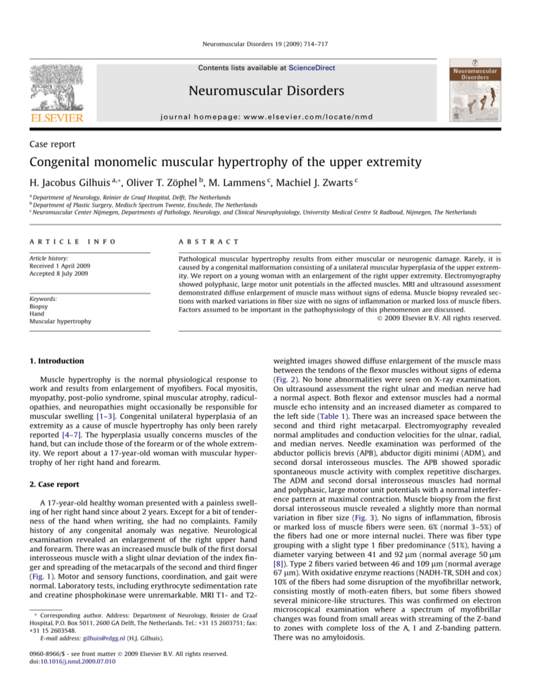
caused by a congenital malformation consisting of a unilateral muscular hyperplasia of the upper extremity. We report on a young woman with an enlargement of the right upper extremity. Electromyography
showed polyphasic, large motor unit potentials in the affected muscles. MRI and ultrasound assessment
demonstrated diffuse enlargement of muscle mass without signs of edema. Muscle biopsy revealed sections with marked variations in fiber size with no signs of inflammation or marked loss of muscle fibers.
Factors assumed to be important in the pathophysiology of this phenomenon are discussed.
Ó 2009 Elsevier B.V. All rights reserved.
1. Introduction
Muscle hypertrophy is the normal physiological response to
work and results from enlargement of myofibers. Focal myositis,
myopathy, post-polio syndrome, spinal muscular atrophy, radiculopathies, and neuropathies might occasionally be responsible for
muscular swelling [1–3]. Congenital unilateral hyperplasia of an
extremity as a cause of muscle hypertrophy has only been rarely
reported [4–7]. The hyperplasia usually concerns muscles of the
hand, but can include those of the forearm or of the whole extremity. We report about a 17-year-old woman with muscular hypertrophy of her right hand and forearm.
2. Case report
A 17-year-old healthy woman presented with a painless swelling of her right hand since about 2 years. Except for a bit of tenderness of the hand when writing, she had no complaints. Family
history of any congenital anomaly was negative. Neurological
examination revealed an enlargement of the right upper hand
and forearm. There was an increased muscle bulk of the first dorsal
interosseous muscle with a slight ulnar deviation of the index finger and spreading of the metacarpals of the second and third finger
(Fig. 1). Motor and sensory functions, coordination, and gait were
normal. Laboratory tests, including erythrocyte sedimentation rate
and creatine phosphokinase were unremarkable. MRI T1- and T2* Corresponding author. Address: Department of Neurology, Reinier de Graaf
Hospital, P.O. Box 5011, 2600 GA Delft, The Netherlands. Tel.: +31 15 2603751; fax:
+31 15 2603548.
E-mail address: gilhuis@rdgg.nl (H.J. Gilhuis).
0960-8966/$ - see front matter Ó 2009 Elsevier B.V. All rights reserved.
doi:10.1016/j.nmd.2009.07.010
weighted images showed diffuse enlargement of the muscle mass
between the tendons of the flexor muscles without signs of edema
(Fig. 2). No bone abnormalities were seen on X-ray examination.
On ultrasound assessment the right ulnar and median nerve had
a normal aspect. Both flexor and extensor muscles had a normal
muscle echo intensity and an increased diameter as compared to
the left side (Table 1). There was an increased space between the
second and third right metacarpal. Electromyography revealed
normal amplitudes and conduction velocities for the ulnar, radial,
and median nerves. Needle examination was performed of the
abductor pollicis brevis (APB), abductor digiti minimi (ADM), and
second dorsal interosseous muscles. The APB showed sporadic
spontaneous muscle activity with complex repetitive discharges.
The ADM and second dorsal interosseous muscles had normal
and polyphasic, large motor unit potentials with a normal interference pattern at maximal contraction. Muscle biopsy from the first
dorsal interosseous muscle revealed a slightly more than normal
variation in fiber size (Fig. 3). No signs of inflammation, fibrosis
or marked loss of muscle fibers were seen. 6% (normal 3–5%) of
the fibers had one or more internal nuclei. There was fiber type
grouping with a slight type 1 fiber predominance (51%), having a
diameter varying between 41 and 92 lm (normal average 50 lm
[8]). Type 2 fibers varied between 46 and 109 lm (normal average
67 lm). With oxidative enzyme reactions (NADH-TR, SDH and cox)
10% of the fibers had some disruption of the myofibrillar network,
consisting mostly of moth-eaten fibers, but some fibers showed
several minicore-like structures. This was confirmed on electron
microscopical examination where a spectrum of myofibrillar
changes was found from small areas with streaming of the Z-band
to zones with complete loss of the A, I and Z-banding pattern.
There was no amyloidosis.
H.J. Gilhuis et al. / Neuromuscular Disorders 19 (2009) 714–717
715
Fig. 1. (Right hand) Notice the increased muscle bulk of the first dorsal interosseous muscle, the ulnar deviation of the index finger, and the spread of the second and third
metacarpals.
Fig. 2. MRI T1-weighted image of the both hands revealing diffuse enlargement of the muscles of the right hand.
716
H.J. Gilhuis et al. / Neuromuscular Disorders 19 (2009) 714–717
Table 1
Echo of both forearms.
Muscles
Side
Echo intensity
z-Score
Diameter
z-Score
Flexor forearm
Flexor forearm
Extensor forearm
Extensor forearm
Right
Left
Right
Left
44
36
37
41
0.0
1.3
0.0
0.0
2.76
2.11
1.52
1.28
2.2
0.5
0.0
0.0
Fig. 3. (A) Muscle biopsy with variation of fiber caliber and fibers with internal
nuclei (HE, bar = 100 lm). (B) Moth-eaten fibers and some fibers with small corelike structures (SDH, bar = 100 lm).
The clinical symptoms as well as the MRI abnormalities remained unchanged during a follow-up of 2 years.
3. Discussion
Muscular hyperplasia due to a congenital unilateral upper-limb
hypertrophy is a rare phenomenon. So far, only 11 clearly defined
cases have been described in the German and English medical literature [4–7] An additional eight cases have been reported in the
Japanese literature [7].The age of these patients from the German
and English literature ranged from 2 to 19 years. None of them
had a progressive course. Of these cases, three had only involvement of the hand, two of the hand and forearm, whereas the others
had involvement of the whole upper extremity, including the
shoulder. All patients, including the present case, had an ulnar
deviation of the fingers and an increased spread at the metacarpal
joints. None of them had a positive family history nor had any
other congenital abnormalities. Except for hypertrophy of the muscles, nine patients have aberrant muscles. So far, in only two other
cases histological examination of muscle tissue was performed
[6,7]. These biopsies did not reveal any abnormalities. In contrast,
the present case showed myopathic changes with especially alterations of the intermyofibrillar network.
Congenital unilateral muscular hyperplasia of an extremity is
distinctly different from other causes of hand or upper-limb hypertrophy. Proteus syndrome for example, a complex disorder comprising malformations and overgrowth, is characterized by a
progressive course of the abnormalities. In most cases, it is not confined to one extremity [9]. The windblown hand is a (usually) bilateral flexion and adduction contracture of the thumb and narrowing
of the first web space. In addition, there is a flexion contracture and
ulnar deviation of the fingers at the metacarpal joints [6]. Freeman–Sheldon syndrome, a type of distal arthrogryposis, involves
two or more features of distal arthrogryposis: microstomia, whistling-face, nasolabial creases, and ‘H-shaped’ chin dimple [10].
The fourth category, which can give hyperplasia of a limb, are
nerve hamartomas. However, they cause progressive growth, and
were excluded by MRI [6]. Finally, muscle hypertrophy following
nerve injury, so called neurogenic pseudo hypertrophy, is electromyographically characterized by excessive spontaneous discharges
and early recruitment of large motor unit potentials at a high firing
rate. The hypertrophy is restricted to muscles innervated by the
damaged nerve. Biopsy findings in these cases show small groups
of types I and II atrophic muscle fibers and abundant hypertrophic
fibers of mostly type II [2]. Congenital muscular hyperplasia has
never been described in the lower extremities.
The exact etiology of unilateral hyperplasia of the upper-limb remains undetermined. The wide range of age at presentation is probably due to the fact that some patients were not seriously hindered
in daily life. Takka et al. [6] claimed that a changed tendon to muscle
ratio and the increased number of neuromuscular junctions involved cause the hypertrophy of the muscles in these patients. Aberrant muscles, they suggest, might be the unused atrophied muscles
during the evolutionary change of the hand like the Palmaris brevis
muscle. Tanabe et al. [7] assumed that in their case, the deformity of
the hand was due to relaxation of the transverse metacarpal ligaments causing increased spread of the fingers. Imbalance of the
intrinsic muscles of the hand would have caused crossing of the index and middle finger. Although in the present case, the fiber type
grouping suggested a neurogenic origin, the hypertrophy was not
confined to a muscle group innervated by a specific peripheral nerve,
part of the brachial plexus, or nerve root. In addition, repeated MRIs
showed normal intensity of muscle tissue.
Treatment is difficult because of the complexity of the clinical
features. It consists of excision of aberrant and hypertrophic muscles for cosmetic reasons or to correct contractures, but is often not
satisfactory [4,7]. Maintenance of the proportional balance of
intrinsic and extrinsic muscles can become difficult [6]. Pillukat
et al. recommend an operation as early as possible to prevent contractures [4]. In the present case, we opted for a ‘wait and see’ policy, as she was not severely hindered in her daily life, and there
were no cosmetic problems.
Acknowledgement
The authors thank Mrs. F. Oostrom for her secretarial support.
References
[1] Guttmann L. AAEM minimonograph #46: neurogenic muscle hypertrophy.
Muscle & Nerve 1996;19:811–9.
[2] Mattle HP, Hess CW, Ludin HP, Mumenthaler M. Isolated muscle hypertrophy
as a sign of radicular or peripheral nerve injury. J Neurol Neurosurg Psychiatry
1991;54:325–9.
[3] Zabel JP, Peutot A, Chapuis D, Batch T, Lecocq J, Blum A. Neurogenic muscle
hypertrophy: imaging features in three cases and review of the literature. J
Radiol 2005;86:133–41 [in French].
H.J. Gilhuis et al. / Neuromuscular Disorders 19 (2009) 714–717
[4] Pillukat T, Lanz U. Congenital unilateral muscular hyperplasia of the hand – a
rare malformation. Handchir Mikrochir Plast Chir 2004;36:170–8 [in German].
[5] So YC. An unusual association of the windblown hand with upper limb
hypertrophy. J Hand Surg 1992;17:113–7.
[6] Takka S, Kazuteru D, Hattory Y, Kitajima I, Sano K. Proposal of new category for
congenital unilateral upper limb muscular hypertrophy. Ann Plast Surg
2005;54:97–102.
[7] Tanabe K, Tada K, Doi T. Unilateral hypertrophy of the upper extremity due to
aberrant muscles. J Hand Surg 1997;22:253–7.
717
[8] Polgar J, Johnson MA, Weightman D, Appleton D. Data on fibre size in
thirty-six
human
muscles.
An
autopsy
study.
J
Neurol
Sci
1973;19:307–18.
[9] Satter E. Proteus syndrome: 2 case reports and a review of the literature. Cutis
2007;297:302.
[10] Stevenson DA, Carey JC, Palumbos J, Rutherford A, Dolcourt J, Bamshad MJ.
Clinical characteristics and natural history of Freeman–Sheldon syndrome.
Pediatrics 2006;117:754–62.


