Root, Stem, Leaf Anatomy Lab Worksheet
advertisement
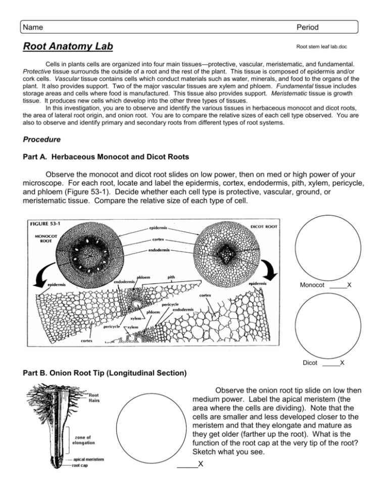
Name Period Root Anatomy Lab Root stem leaf lab.doc Cells in plants cells are organized into four main tissues—protective, vascular, meristematic, and fundamental. Protective tissue surrounds the outside of a root and the rest of the plant. This tissue is composed of epidermis and/or cork cells. Vascular tissue contains cells which conduct materials such as water, minerals, and food to the organs of the plant. It also provides support. Two of the major vascular tissues are xylem and phloem. Fundamental tissue includes storage areas and cells where food is manufactured. This tissue also provides support. Meristematic tissue is growth tissue. It produces new cells which develop into the other three types of tissues. In this investigation, you are to observe and identify the various tissues in herbaceous monocot and dicot roots, the area of lateral root origin, and onion root. You are to compare the relative sizes of each cell type observed. You are also to observe and identify primary and secondary roots from different types of root systems. Procedure Part A. Herbaceous Monocot and Dicot Roots Observe the monocot and dicot root slides on low power, then on med or high power of your microscope. For each root, locate and label the epidermis, cortex, endodermis, pith, xylem, pericycle, and phloem (Figure 53-1). Decide whether each cell type is protective, vascular, ground, or meristematic tissue. Compare the relative size of each type of cell. Monocot _____X Dicot _____X Part B. Onion Root Tip (Longitudinal Section) Observe the onion root tip slide on low then medium power. Label the apical meristem (the area where the cells are dividing). Note that the cells are smaller and less developed closer to the meristem and that they elongate and mature as they get older (farther up the root). What is the function of the root cap at the very tip of the root? Sketch what you see. _____X Part C. Root Systems Using Figure 53-3, observe the different types of root systems. Formulating Generalizations 1. The blue-stained material in the cortex of the dicot root is starch. Why is it found in the root and how did it get there? 2. How do xylem patterns differ in monocot and dicot roots? 3. What are four basic functions of roots? Stem Anatomy Lab As in roots, protective, vascular, fundamental, and meristematic tissues are found in stems. The difference between roots and stems may be slight or significant. In this investigation, you are to observe and identify the tissues in an herbaceous monocot stem, and herbaceous dicot stem, and a woody stem. Part A. Herbaceous Monocot and Dicot Stems Observe the monocot and dicot slides on low power, then on high power of your microscope. Make a sketch of each stem and label the epidermis, cortex, vascular bundles, xylem, phloem, cambium, and pith (Figure 54-1). Know the functions of each cell type. Decide whether each cell type is protective, vascular, ground, or meristematic tissue. Compare the relative size of each cell to the other cells. Monocot _____X Dicot _____X Part B. Tilia Stem (Woody growth) Observe the Tilia stem slide on low power, then on high power of your microscope. Sketch and label the epidermis, cork, cortex, phloem, cambium, spring wood, summer wood, xylem, pith rays, and pith (Figure 54-2). Know the functions of each cell type. Decide whether each cell type is that of protective, vascular, ground, or meristematic tissue. Compare the relative cell sizes. _____X Part C. Woody Twig Observe the woody twig. Locate and identify the terminal bud, lateral bud, lenticels (small pores in the bark), bud scale, bud scale scar, node, internode, and leaf scar (Figure 54-3). Know the functions of each part of a woody twig. Determine whether the stem has an alternate or opposite leaf arrangement. The area between two bud scale scars is one year’s growth. Count the years of growth on the twig. ______________years old Formulating Generalizations 1. How is woody xylem in Tilia different from xylem in herbaceous monocots and dicots? 2. How do monocot and dicot stems differ in location of cambium, xylem, and phloem? 3. Explain how you would tell if one growing season was more favorable for growth than others. 4. How can you determine the past arrangement of leaves on a bare twig? Leaf Structure And Function Lab Leaves are the main organs of food production and photosynthesis in green plants. In most plants, leaves are generally flattened. The flattened leaves have a large surface area for sun exposure. The flattened part of a leaf is called the blade (Figure 50-1). A leaf is attached to the stem of the plant by a petiole. The petiole contains vessels that carry water to the leaf (xylem) and carry food away from the leaf (phloem). Leaf tissues are grouped into three types: epidermal, mesophyll, and vascular (Figure 50-2). Most leaves have all the tissues shown in Figure 50-2. However, some leaves have extra or fewer structures. Differences in leaf structures are adaptations to the type of environment in which the plant lives. In this investigation, you are to observe prepared microscopic slides of leaf cross sections. You are to locate, identify, and distinguish the various structures. You are to describe the differences of the three types of tissues found in the leaf types. Procedure Using low and high power of your microscope, examine the slides of the leaf cross sections. Label cutin, epidermis, veins (xylem, phloem, and bundle sheath), palisade tissue, spongy tissue, guard cells, and stomata. Make a sketch of each cross section. See Figure 50-2. Part A. Monocot _____X vs. Dicot Leaf (c.s.) _____X 1. On which layer (upper or lower) of epidermis are guard cells found? Why? 2. What is the function of the spongy mesophyll? 3. Which leaf, monocot or dicot, appears to have veins that are more evenly spaced in cross section? Why? Part B. Water Lily (Lily pad) 4. Why are the air spaces in the spongy tissue two or four times larger than those in all other leaves? 5. On which layer of epidermis, upper or lower, are the guard cells and stomata found? Why are stomata found here? _____X Part C. Yucca (desert plant) 6. Why do the guard cells and the stomata appear to be small? 7. Why are air spaces almost entirely absent? Formulating Generalizations _____X 1. Why are palisade cell so tightly packed in the upper surface of the leaf? 2. Why are the bundle sheaths more prominent in some leaves than in others (look back at C3 vs. C4 plants)? 3. What does the thickness of the cutin have to do with the environment in which the plant lives?
