D- Integumentary
advertisement
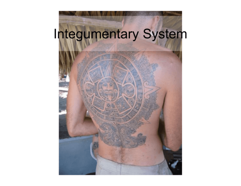
Integumentary System Functions of the integumentary system • Protection from the environment-the skin is the superficial surface of the body • Thermoregulation-secretions from sweat glands in the skin cool the body down • Storage of lipids-adipose tissue (fat) • Vitamin D synthesis • Provides sensory info-sensory receptors located in the skin Fig 4.1 Adipose CT Fig 4.2 Layers of the epidermis Stratum basal (germinativum)-attached to basement membrane, contains stem cells & melanocytes Stratum spinosum-keratinization begins Stratum granulosum-process of adding keratin continues Stratum lucidum-only in thick skin Stratum corneum-at surface of skin Layers of dead interlocking keratinocytes Contains large amount of keratin Makes a dry water resistant layer Epidermis Fig 4.3 Thick & thin skin • Thick skin has 5 layers in the epidermisincludes the stratum lucidum, plantar/palmar • Thick skin has a thicker stratum corneum Fig 4.4 Fingerprints-thick skin • Epidermis-epidermal ridges • Dermis-dermal papillae Fig 4.4 Dermis • • • • • Composed of connective tissue Highly vascular Contain nerves and sensory receptors Located deep to the epidermis Has two layers: – Papillary layer provides nutrients, O2 etc to the epidermis – Reticular layer-interwoven network of collage fibers surrounding dermal organs Papillary & Reticular layers Papillary layer Consists of areolar CT provides nutrients, O2 etc to the epidermis Tattoo ink is injected into the papillary layer Reticular layer – Reticular layer-interwoven network of collage fibers surrounding dermal organs – Wrinkles and stretch marks arise from degradation of the reticular layer Lines of cleavage-clinical aspect • Collagen & elastic fibers are arranged in parallel bundles in the skin • Incisions parallel to the lines of cleavage heal faster than incisions at a right angle to the line of cleavage Hypodermis • Loose Ct with adipose cells • Regional distributions of adipose in males and females • • • • Stabilizes position of organs Reduces heat loss Energy reserve Cushion ?’s about the integument • Text chapter 4 Accessory structures • Hair, nails, & glands in the skin (dermis) • Hair grows everywhere except areas with thick skin and portions of the external genitalia • Hair is formed in organs called hair follicles • Hair give added sensory info and protects orifices of the body (nostrils, ears) Hair • Types of hairs on the body: • Vellus hairs-“peach fuzz” over most of the body • Intermediate hairs-hairs stimulated by hormones-pubic hair, beard, distal appendages • Terminal hairs-hairs on head, eyebrows, eyelashes • Hair is dead keratinized epithelial cells hard keratin follicle Soft keratin hard keratin Fig 4.10 Glands in the skin Fig 4.12 Sebaceous glands • Branch off of hair follicles • Release oily secretion on to hair Fig 4.9 Sweat glands • Apocrine-in the axillary, areolae & inguinal regions • -secrete into hair • Merocrine (eccrine)all over the body – Secrete onto skin – Smaller and more superficial than apocrine glands Fig 4.14 • Mammary glands-modified apocrine glands that release breast milk • Cerumious glands-modified merocrine glands that release cerumen (ear wax) mechanism holocrine of secretion Type of gland merocrine apocrine Sebaceous merocrine Mammary glands (eccrine) & glands Apocrine glands Fig 3.9 Nails • Protect distal ends of finger & toes • Stratum corneum forms the hyponychium and eponychium • Blood vessels give the pink color Fig 4.15 Layers of the integument-review superficial • Epidermis-stratified squamous epithelial tissue – Stratum corneum-thicker in thick skin-palmar/plantar – Stratum lucidum-only in thick skin – Stratum granulosum-contains keratin & (melanin in people of African decent) – Stratum spinosum-contains melanin & keratinocytes – Stratum basal (germinativum)-contain melanocytes-melanin • Dermis - Papillary layer-areolar CT deep superficial • Dermis – Papillary layer- areolar CT • Eccrine sweat glands-watery secretions • Sebaceous glands- oily secertions • Meissners corpuscle-sensory receptors for soft touch – Reticular layer- dense irregular CT • Apocrine sweat glands- smelly secretions • Hypodermis – Adipose CT – Pacinian corpuscles-sensory receptors for deep pressure deep Adipose CT Fig 4.2 Burns to the skin classification damage Affected organs Appearance and sensation 1st degree burn Superficial cells of the epidermis are killed. Dermis cells are injured-papillary layer Hair follicles & glands unaffected Inflamed, tender 2nd degree burn Injury to dermisreticular layer Hair follicles & glands may be affected Blister, pain 3rd degree burn All dermal cells are killed. Injury to the hypodermis Sensory nerves, accessory structure, blood vessels destroyed Charred, less pain than 1st and 2nd FYI Aging & the Integumentary system Changes that occur Result Epidermis thins-less More prone to injury/infection germinative cell activity Decreased # of Langerhans Reduced immune function cells Decreased melanocyte More sensitivity to activity sun/sunburn Reduced Vitamin. D synthesis Muscle/bone weakness Decreased dermal blood Reduced ability to regulate supply & sweat/oil gland temperature, dryer skin activity Hair follicles function Thinner hairs, grey/white decreases hairs, balding Dermis thins, elastic fiber Weaker sagging wrinkled skin network shrinks Skin repairs slowly Recurring infections • Photos of models • http://www.rwc.uc.edu/ap/aphome.htm Lab 5 Adipose CT Fig 4.2 Fig 4.9

