Muscles How muscles contract
advertisement
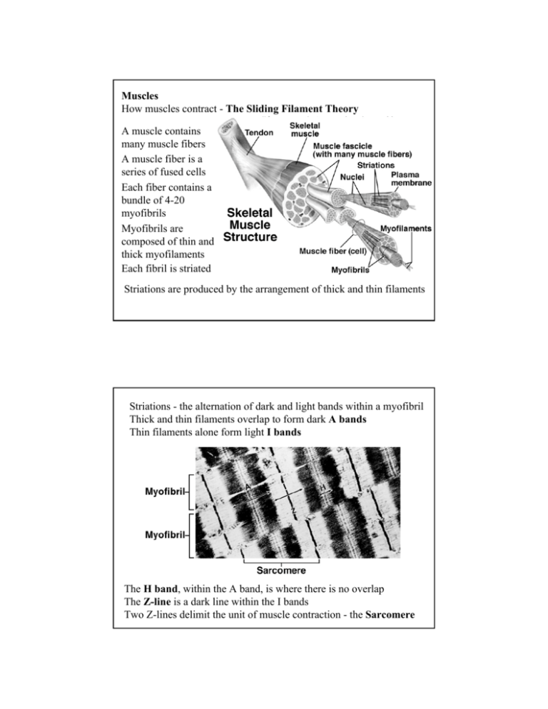
Muscles How muscles contract - The Sliding Filament Theory A muscle contains many muscle fibers A muscle fiber is a series of fused cells Each fiber contains a bundle of 4-20 myofibrils Myofibrils are composed of thin and thick myofilaments Each fibril is striated Striations are produced by the arrangement of thick and thin filaments Striations - the alternation of dark and light bands within a myofibril Thick and thin filaments overlap to form dark A bands Thin filaments alone form light I bands The H band, within the A band, is where there is no overlap The Z-line is a dark line within the I bands Two Z-lines delimit the unit of muscle contraction - the Sarcomere During muscle contraction, sarcomeres shorten, the distance between Z-lines decreases - the width of the H and I-bands also decreases Thick filaments are composed of the protein myosin Thin filaments are composed of the protein actin relaxed contracted During contraction the amount of overlap between actin and myosin increases Contraction is produced by an interaction between actin and myosin Through the formation of cross bridges, the actin is pulled into the space between the myosin filaments Cross-bridges extend from thick to thin filaments Each myosin molecule has a protruding head Heads form cross-bridges by interacting with actin Thick filaments are composed of many myosin molecules Thin filaments are composed of a double helix of globular actin molecules During contraction, myosin heads bind to actin molecules of the thin filaments Flexing of the myosin heads pulls the thin filaments over the thick filaments - shortening the sarcomere Muscle contraction requires ATP Cleavage of ATP activates the myosin head When activated, the myosin head can form a cross-bridge to actin After binding to actin, ADP is released from myosin head head flexes, pulling the actin filament over myosin ATP is required to detach myosin from actin and repeat cycle Multiple rounds of the cycle at many myosin heads results in shortening of the sarcomere ATP is normally present in live muscle at all times - the depletion of ATP in dying muscle causes formation of permanent cross-bridges the dying muscle becomes rigid - “rigor mortis” Muscle contraction requires Ca++ - Ca++ presence exposes the actin binding sites through an interaction between Ca and troponin Tropomyosin associates with actin and blocks binding sites on actin when Ca++ is absent Troponin associates with tropomyosin and causes conformational change in tropomyosin when Ca++ associates with troponin When muscles are relaxed Ca++ is stored in sarcoplasmic reticulum release causes contraction cycle to begin The sarcoplasmic reticulum (SR) surrounds each myofibril, and is connected to the muscle cell membrane (sarcolemma) by T-tubules Skeletal muscle fiber contraction is the result of nervous stimulation Nerve stimulation results in change in permeability of the sarcolemma and SR The stimulated SR membrane becomes permeable to Ca++ releasing stored Ca++ - and the contraction cycle can begin Nerves connect to muscles at neuromuscular junctions - a single nerve may connect with more than one muscle fiber The nerve and the muscle fibers it innervates is a “motor unit” Nerve stimulation involves the release of a neurotransmitter from the axon terminus at the neuromuscular junction - “excitationcontraction coupling” The neurotransmitter is Acetylcholine (ACh) ACh binds to receptor proteins on the sarcolemma and changes membrane permeability - change is transmitted along membranes of the Ttubules to the SR Relaxation of muscle fibers involves the uptake of Ca++ from the cytoplasm of the muscle fiber by the SR Recruitment: Small force muscle contractions involve few motor units - Higher force contractions involve an increased number of motor units Types of Muscle Fibers Fast-twitch fibers (type II fibers) - common in muscles that move eyes - reach maximum tension in 7.3 msec Slow-twitch fibers (type I fibers) - common in leg muscle - reach maximum tension in 100 msec Many muscles have a mixture of slow and fasttwitch fibers Slow-twitch fibers are able to sustain contractions over long periods without fatigue use aerobic respiration, have rich vascular supply, many mitochondria, high concentration of myoglobin gives them a red color, “red fibers” Fast-twitch fibers are liable to fatigue use anaerobic respiration, have stored glycogen have reduced vascular supply, fewer mitochondria, less myoglobin, “white fibers” Muscle fatigue is associated with the build-up of lactic acid due to anaerobic metabolism of glucose The production of lactic acid allows glycolysis to continue for short periods in the absence of oxygen but eventually lactic acid buildup inhibits enzymes of glycolysis Lactic acid build-up produces an “oxygen debt” Lactic acid must be removed from muscle by aerobic metabolism after strenuous activity ceases Muscle Fiber Twitch - A single brief contraction a single impulse on motor neuron produces a single twitch contraction followed by rapid and relaxation increasing stimulation can increase the strength of a twitch up to a maximum If two impulses are applied in rapid succession a greater state of contraction results - second twitch adds to first - “summation” A series of rapid impulses applied at increasing frequency produces a smooth sustained contraction - “tetanus” - as in normal muscle contraction Cardiac and Smooth Muscle can both contract spontaneously Cardiac muscle is striated - but muscle cells are not arranged as in skeletal muscles Muscle cells are branched and connected to each other at intercalated disks - have gap at disks that allow stimulation to be quickly passed from cell to cell Stimulation begins at “pacemaker” cells Smooth Muscle - surrounds hollow organs - stomach, intestine, arteries, bladder, uterus Capable of sustained contractions Thick and thin filaments lie parallel to each other but groups are arranged irregularly - anchored to cell membrane or dense bodies Lack SR - Ca++ enters from extracellular fluid - Ca++ binds calmodulin - complex activates enzyme that phosphorylates myosin heads - allows cross-bridge formation variation in Ca++ concentration varies strength of contraction Some smooth muscles do contract in response to nervous stimulation - e.g. muscles of iris Other smooth muscles contract spontaneously - through the action of stimulatory cells within the muscle - e.g. gut Smooth muscle is capable of contraction after extreme stretching not true of striated muscle


