From www.bloodjournal.org by guest on March 4, 2016. For personal use only.
High Risk of Thrombosis in Patients Homozygous for Factor
(Activated Protein C Resistance)
V Leiden
By F.R. Rosendaal, T. Koster, J.P. Vandenbroucke, and P.H. Reitsma
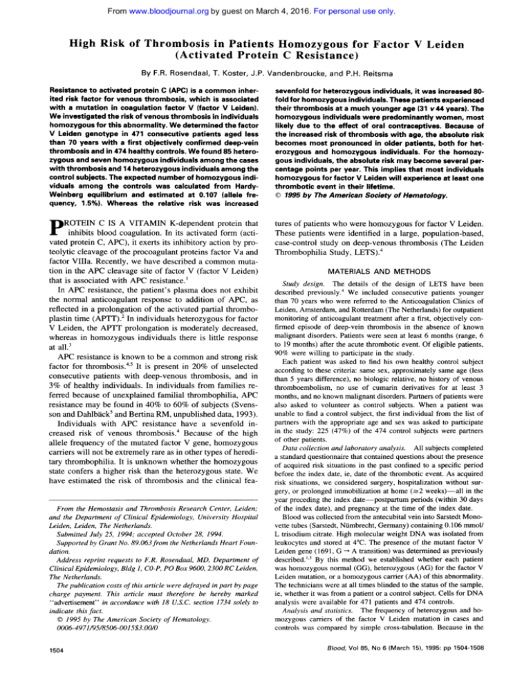
Resistance to activated protein C (APC) is a common inherited risk factor for venous thrombosis, which is associated
with a mutation in coagulation factor V (factor V Leiden).
We investigated the risk ofvenous thrombosis in individuals
homozygous for this abnormality. We determined the factor
V Leiden genotype in 471 consecutive patients aged less
than 70 years with a first objectively confirmed deep-vein
thrombosis and in 474 healthy controls. We found 85 heterozygous and seven homozygous individuals among the cases
with thrombosis and 14 heterozygous individualsamong the
control subjects. The expected number of homozygous individuals among the controls was calculated from HardyWeinberg equilibrium and estimated at 0.107 (allele frequency, 1.5%). Whereas the relative risk was increased
sevenfold for heterozygous individuals, it was increased 80fold for homozygous individuals.These patients experienced
their thrombosis at a much younger age (31 v44 years). The
homozygous individuals were predominantly women, most
likely due to the effect of oral contraceptives. Because of
the increased risk ofthrombosis with age, the absolute risk
becomes most pronounced in older patients, both for heterozygous and homozygous individuals. For the homozygous individuals, the absolute risk may become several percentage points per year. This implies that most individuals
homozygous for factor V Leiden will experience at least one
thrombotic event in their lifetime.
0 1995 by The AmericanSociety of Hematology.
P
tures of patients who were homozygous for factor V Leiden.
These patients were identified in a large, population-based,
case-control study on deep-venous thrombosis (The Leiden
Thrombophilia Study, LETS).4
ROTEIN C IS A VITAMIN K-dependent protein that
inhibits blood coagulation. In its activated form (activated protein C, APC), it exerts its inhibitory action by proteolytic cleavage of the procoagulant proteins factor Va and
factor VIIIa. Recently, we have described a common mutation in the APC cleavage site of factor V (factor V Leiden)
that is associated with APC resistance.’
In APC resistance, the patient’s plasma does not exhibit
the normal anticoagulant response to addition of APC, as
reflected in a prolongation of the activated partial thromboplastin time (APTT).*In individuals heterozygous for factor
V Leiden, the AP’IT prolongation is moderately decreased,
whereas in homozygous individuals there is little response
at all.’
APC resistance is known to be a common and strong risk
factor for thromb~sis.~.~
It is present in 20% of unselected
consecutive patients with deep-venous thrombosis, and in
3% of healthy individuals. In individuals from families referred because of unexplained familial thrombophilia, APC
resistance may be found in 40% to 60% of subjects (Svensson and Dahlback5and Bertina R M , unpublished data, 1993).
Individuals withAPC resistance have a sevenfold increased risk of venous thromb~sis.~
Because of thehigh
allele frequency of the mutated factor V gene, homozygous
carriers will not be extremely rare as in other types of hereditary thrombophilia. It is unknown whether the homozygous
state confers a higher risk than the heterozygous state. We
have estimated the risk of thrombosis and the clinical fea-
From the Hemostasis and Thrombosis Research Center, Leiden;
and the Department of Clinical Epidemiology, Universily Hospital
Leiden, Leiden, The Netherlands.
Submitted July 25, 1994; accepted October 28, 1994.
Supported by Grant No. 89.063from the Netherlands Heart Foundation.
Address reprint requests to F.R. Rosendaal, MD, Department of
Clinical Epidemiology, Bldg I , CO-P, PO Box 9600,2300 RC Leiden,
The Netherlands.
The publication costs ofthis article were defrayed
in part by page
chargepayment. This article must therefore be hereby marked
“advertisement” in accordance with 18 U.S.C. section 1734 solely to
indicate this fact.
0 1995 by The American Society of Hematology.
0006-4971/95/8506-0015$3.00/0
1504
MATERIALS ANDMETHODS
Study design. The details of the design ofLETS have been
described previously? We included consecutive patients younger
than 70 years who were referred to the Anticoagulation Clinics of
Leiden, Amsterdam, and Rotterdam (The Netherlands) for outpatient
monitoring of anticoagulant treatment after a first, objectively confirmed episode of deep-vein thrombosis in the absence of known
malignant disorders. Patients were seen at least 6 months (range, 6
to 19 months) after the acute thrombotic event. Of eligible patients,
90% were willing to participate in the study.
Each patient was asked to find his own healthy control subject
according to these criteria: same sex, approximately same age (less
than 5 years difference), no biologic relative, no history of venous
thromboembolism, no use of cumarin derivatives for at least 3
months, and no knownmalignant disorders. Partners of patients were
also asked to volunteer as control subjects. When a patient was
unable to find a control subject, the first individual from the list of
partners with the appropriate age and sex was asked to participate
in the study: 225 (47%) of the 474 control subjects were partners
of other patients.
Data collection and laboratoty analysis. All subjects completed
a standard questionnaire that contained questions about the presence
of acquired risk situations in the past confined to a specific period
before the index date, ie, date of the thrombotic event. As acquired
risk situations, we considered surgery, hospitalization without surgery, or prolonged immobilization at home ( 2 2 weeks)-all in the
year preceding the index date-postpartum periods (within 30 days
of the index date), and pregnancy at the time of the index date.
Blood wascollected from the antecubital vein into Sarstedt Monovette tubes (Sarstedt, Numbrecht, Germany) containing 0.106 m o l /
L trisodium citrate. High molecular weight DNA was isolated from
leukocytes and stored at 4°C. The presence of the mutant factor V
Leiden gene (1691, G + A transition) was determined as previously
de~cribed.’.~
By thismethodwe established whether each patient
was homozygous normal (GG),heterozygous (AG) for the factor V
Leiden mutation, or a homozygous camer (AA) of this abnormality.
The technicians were at all times blinded to the status of the sample,
ie, whether it was from a patient or a control subject. Cells for DNA
analysis were available for 471 patients and 474 controls.
Anulysis and statistics. The frequency of heterozygous and homozygous carriers of the factor V Leiden mutation in cases and
controls was compared by simple cross-tabulation. Because in the
Blood, Vol 85, No 6 (March 15). 1995: pp 1504-1508
From www.bloodjournal.org by guest on March 4, 2016. For personal use only.
1505
HOMOZYGOUS FACTOR V LEIDEN AND THROMBOSIS
analysis of the risk associated with the heterozygous state, sex and
age did not appear to be confounding variables (as they were not
expected to be for autosomal genetic abnormalities), relative risk
estimates for the heterozygous state were obtained by calculation of
unmatched exposure odds ratios. A 95% confidence interval (CI95%)
was constructed according to Woolf.6
The risk associated with the homozygous state could not be estimated in this standard fashion, as no homozygous individuals were
found among the controls. Therefore, under the assumption of
Hardy-Weinberg equilibrium (in the controls), the expected number
of homozygous individuals in a control population was calculated,
and the odds ratio was subsequently estimated in the standard fashion. The variance of the (log) odds ratio for the homozygous state
was estimated by a modification of the method of Woolf.6 When
each cell of the two-by-two table with cell contents a, b, c, and d
is considered tobe the realization of a Poisson distribution, the
variance of the log(0R) is I/a + 1/b + l/c + l/d (as var[ln(x)] =
I/x).~
When the number of individuals with GG and AA genotypes
are counted in the cases and calculated from Hardy-Weinberg equilibrium for the controls, which requires a quadratic transformation,
the odds ratio can be estimated as AA cases/GG cases divided by
(A controls/G controls)’, in which AA and GG are the number of
genotypes (individuals), and A and G the number of alleles; the
expected number of homozygous controls is (A controls)’/Z(G controls). Thus, the In(0R) is ln(AA cases) - In(GG cases) - 21n(A
controls) + 21n(G controls). The variance can now be estimated by
assuming that all parameters have a Poisson distribution, which (as
var[2ln(x)] = 2’/x) leads to var[ln(OR)] = ]/AA + I I G G + 4/G +
4/A.
The absolute risk for thrombosis for the various genotypes and
ages was calculated by first partitioning the total number of person
years in the origin population (as derived from census information
from the municipal authorities) under the assumption of HardyWeinberg equilibrium. Dividing the cases in each subgroup (genotype, age) by these person years leads to estimates of the absolute
risks. Subsequently, these crude incidence data were modelled after
logarithmic transformation in a weighted least square regression
model, with three age classes (0 to 29, 25 years; 30 to 49,40 years;
50 to 69, 60 years), indicator variables for the heterozygous (0, 1)
and the homozygous state (0, l), weighted for the number of cases
in each stratum. This method, in which stratum-specific incidence
rates are first estimated and then smoothed (smooth-last’) by
weighted least square regression, has been described by Grizzle et
al.’ Because for a Poisson distribution the variance of the number
of cases equals the number of cases, this closely resembles fitting
of a Poisson regression model. The model will lead to more stable
estimates than the crude incidence figures, especially for the homozygous state, under the assumption that the incidence rate ratio for
the homozygous state (and the heterozygous state) is constant over
the age strata [for the log(incidence rate), log(X)]. This model can
be written as: log@) = a + p, * age + * AG(0, 1 ) + p, * AA@,
l), which can subsequently be used to calculate estimates for the
absolute risk (by entering all covariate values and the estimated
coefficients in this equation) and for the relative risk (as the antilogarithm of the coefficients).
RESULTS
Among 47 1 patients, we found 85 (18%)who were heterozygous and seven (1.5%) who were homozygous for the
defect, whereas the other 379 (80%) did not carry the factor
V Leiden mutation. Among the 474 controls, 14 (2.9%) were
heterozygous, and all other 460 were normal; there were no
homozygous individuals among the controls.
The homozygous individuals experienced thrombosis at a
markedly younger age than the other patients: the median
Table 1. General Characteristics of 471 Thrombosis Patients by
Factor V Genotype
n
Age ( v s )
Median
Range
Sex
No. of men (%)
No. of women (%)
GG
AG
AA
379
85
7
46
15-69
44
17-69
31
22-55
162 (43)(14) 1 (46)39
217 (57)
(86)
466 (54)
age at thrombosis was 31 years compared with 44 years in
the heterozygous and 46 years in the patients without the
mutation (Table 1).
The clinical course of the deep vein thrombosis in the
homozygous patients was unremarkable (see Appendix). All
suffered from deep venous thrombosis of the proximal deep
veins of the leg. Four were hospitalized for heparinization,
and three were treated as outpatients with cumarin derivatives only, according to the regional treatment policies for
venous thrombosis.
Six (86%) of the seven homozygous patients were women,
compared with 46 (54%) of the heterozygous and 217 (57%)
of the individuals without the mutation (Table 2). Also, six
of these seven patients had blood group A, compared with
249 (54%) of the other 464 cases. Of the five homozygous
women aged 45 years and younger, three used oral contraceptives at the time of the thrombotic event, which was
similar to the use of oral contraceptives in all cases (105 of
159, 66%).
In five (71%) of the seven homozygous patients, the
thrombosis apparently had occurred spontaneously, and in
two there had been a predisposing factor for thrombosis in
the year preceding the event (one had hip surgery 20 days
before the thrombosis, and one hadbeen admitted to the
hospital overnight after giving birth 60 days before the
thrombotic event; see Table 2). Among the 85 heterozygous
individuals, an acquired risk factor had been present in 25
(29%) patients, and among the normal (GG) patients, in 131
(35%) of 379.
Previous risk situations (operations, pregnancies, hospital
admissions) without thrombotic consequences were less frequent in the patients homozygous for factor V Leiden than
in the other patients. Still, five of the seven homozygous
patients had encountered risk situations in the past without
a subsequent thrombosis (two hadhad surgery, four had
given birth to five children).
The seven patients were observed for an average of 2
years without long-term oral anticoagulation after the first
thrombotic event. One patient had a recurrent thrombosis (1
in 13.4 years, 7.4% per year recurrence risk for these seven
patients). Of the 14 parents of these seven patients, three
had a history of venous thrombosis, which is approximately
five times higher than expected.’
Under Hardy-Weinberg equilibrium, the relative frequency of norma1s:heterozygotes:homozygotesis p2:2pq:q2,
where p is the allele frequency of the normal gene and q of
the abnormal gene. As p2:2pqwas 4601474: 141474,it follows
that the allele frequency of factor V Leiden (4) is 0.015. The
From www.bloodjournal.org by guest on March 4, 2016. For personal use only.
ROSENDAAL ET AL
1506
Table 2. Detailed Characteristics of Seven Homozygous Patients
Patient
No.
Sex (yrs)
90
124
266
173
583
589
944
F
F
F
1.20
APC-SR
Age
55
24
30
22
44
42
31
F
F
F
M
Blood
Group
1.13
1.14
A
A
A
1.14
1.23
1.21
1.19
(male)
occ*
N§
N
N
YIl (60)
Y
N
N
Y
N
Y
0
A
A
A
Predisposing Factorst
(days between risk
situation and VT)
No. of Parents
With History of VT
Arterial
Disease*
0
0
0
0
Y
N
N
N
N
N
N
1
1
1
Y' (20)
N
N
Abbreviations: APC-SR. APC sensitivity ratio; OCC, oral contraceptives; V
T
, venous thrombosis.
* Use of oral contraceptives in the month preceding the thrombosis.
t Surgery, hospital admission, immobilization in the year preceding the thrombosis, childbirth 1 month before the thrombosis, pregnancy at
the time of the thrombosis.
*Angina pectoris, previous myocardial infarction, stroke, or peripheral arterial disease.
5 Menopausal, no use of estrogens.
l 1 Overnight hospital stay after giving birth.
7 Hip surgery.
allele frequencies of p = 0.985 and q = 0.015 conform to
a distribution among 474 unselected individuals of 459.9
(GG), 14.0 (AG), and 0.107 (AA).
The expected number of homozygous individuals (S2) of
0.107 among 474 controls leads to an odds ratio for the
homozygous state of (7/379)/(0.107/460) = 79.4. So, the
risk of thrombosis for homozygous individuals is almost 80
times increased compared with normal individuals (CI95%,
22 to 289).
Table 3 shows the odds ratio for the three age groups
when this allele frequency is used to calculate the expected
number of homozygous controls ineach age group. Itis
evident that the high relative risk of thrombosis in homozygous individuals diminishes with advancing age. This is in
contrast to the relative risk for heterozygous individuals,
which is more or less constant over age.
Subsequently, we calculated absolute risks (incidence
rates) for the different age groups and genotypes by using
data on the age-distribution in the origin population, and
under the assumption of Hardy-Weinberg equilibrium. As is
shown in Table 3, the incidence increases from only 0.6 per
10,000per year in the youngest age group with GG genotype,
to 16.4 per 10,000 per year for heterozygous individuals in
the older age groups. It is also clear from the ratios in Table
3 that at all age groups the risk for homozygous individuals
is much higher than for heterozygous individuals (78 to 176
per 10,000 per year). However, as these figures are based
on only seven individuals divided over three age groups,
the estimates are unstable andshow an unexpected lower
incidence in the oldest age group. The regression model we
used smooths these estimates, as it assumes a constant relative risk over the age groups. As Fig 1 shows, this model
fits excellently for the normal and heterozygous individuals
(coefficients: constant, -10.06; age, .0293; AG, 1.96; AA,
4.52). The smoothed incidence estimates for homozygous
individuals now increase from 82 per 10,000 person-years
in those aged under 30 years to 227 per 10,000 patient-years
for those aged 50 to 69 years (Fig 1). These estimates imply
that most homozygous patients will experience at least one
thrombotic event in their lifetime.
DISCUSSION
Resistance to APC is a common abnormality, with an
allele frequency for the mutant factor V gene of about 1.5%.
Three percent of the population will be heterozygous, and
homozygous individuals can be expected with a prevalence
of about 2 per 10,000 births.
In this study we show that homozygous individuals have
a high risk of thrombosis, which is considerably higher than
the risk in heterozygous individuals. This conclusion is sup-
Table 3. Odds Ratios andAbsolute Risk of First Thrombosis by Age
Genotype
Incidence Rates/lO" yrs"
Age ( v s )
0-29
30-49
50+
Patients
IGG/AG)
(GGIAGIAA)
Controls
7013 6111712
(1.8-23) 6.5
(9.5-2.049)
140
2 1716
17613514
17315
14213311
(2.2-428) 30
EA*
ORnn (C195%)
,0164
,0502 (3.0-17) 98
7.2(15-652)
110.0 16.4
,0400
ORAG
(C195%)
Personyears
1,134,681
1,006,733
GG
AG
AA
0.678.4
5.1
1.8 176.2 11.8
2.1 682,939
8.0 (3.1-21)
Abbreviations: EM, expected number of homozygous individuals among controls based on overall Hardy-Weinberg equilibrium
q = .015); ORM, odds ratio for homozygous individuals ( v homozygous normals); ORaG, odds ratio for heterozygous
(4' * ncontro,.,nage.Jtrarum,
individuals ( v homozygous normals).
* Crude incidence rates, based on the number of observed cases over the number of patient-years, partitioned according to Hardy-Weinberg
equilibrium (q = ,015).
From www.bloodjournal.org by guest on March 4, 2016. For personal use only.
1507
HOMOZYGOUS FACTOR V LEIDEN AND THROMBOSIS
incidence
30
25
20
l
+
15
10
5
0
0-29
age
Fig1.Crudeandsmoothedincidencerateestimatesper
10"
years for factor V Leiden genotypesby age. The lowed line shows
the estimates for the GG genotype, and the upper line for the AG
genotype (in the full figure). The inserted figure also shows
the estimates for the AA genotype (homozygous factorV Leiden). crude
incidenceestimates; 0, smoothed rates. The smoothedincidence
rates per 10,OOO person years were as follows: GG: 0.9 (0-29 years),
1.4 (30-49 years). and 2.5 (50-69 years); AG. 6.3 ( 0 - 2 9 years), 9.8 (3049 years), 17.6
( 5 0 - 6 9 years); a 81.5 (0-29 years). 126.5 (30-49 years),
and 227.3 (50-69 years).
+,
ported by the young age at which the homozygous individuals experience their first thrombotic event.
It is clear that the risk of thrombosis in homozygous factor
V Leiden is nowhere near the risk of thrombosis in homozygous protein C or protein S deficiency; these abnormalities
lead to neonatal purpura fulminans.l0.'' All of the individuals
with homozygous factor V Leiden lived until adulthood before the first thrombotic event, and one even until late middle
age. Most of the homozygous individuals had experienced
risk situations in the past without thrombosis; for most of
these situations (pregnancy, puerperium), no anticoagulant
prophylaxis will have been prescribed. This shows that APC
resistance should be seen as a quantitative defect (decreased
inactivation rate of factor Va) rather than a qualitative defect
(no protein C activity), as in homozygous protein C deficiency.
A remarkable finding in this study was the predominance
of women among the homozygous patients. Among these
homozygous women, the use of oral contraceptives was as
prevalent as among the other female thrombosis patients.
From this, it follows that the relative risk associated with
oral contraceptive use is the same for homozygous women
as for women with the other genotypes (under the reasonable
assumption that among the healthy control subjects oral contraceptive use is not associated with the factor V genotype:
the exposure odds among the cases divided by the exposure
odds of the controls-the odds ratio-will be the same).
This implies that the already greatly increased risk of a homozygous woman is again multiplied four- to sixfold when
she uses the pill," and so the absolute increase in risk due
to oral contraceptives is much larger in women homozygous
for the factor V Leiden mutation than for other women.I3
This shows that oral contraceptives have the same synergistic
effect with the homozygous factor V Leiden mutation as
we have recently reported for the heterozygous state." The
incidence rates of Table 3 and Fig 1 are overall data, and
so the rates will be somewhat lower for those who do not
use oral contraceptives, at 50 to 100 per 10,000 person years
in the homozygous individuals. These rates clearly demonstrate the different effect of oral contraceptives for the various genotypes: for the youngest women with the GG genotype, use of oral contraceptives with a relative risk of 4 will
only lead to 1 or 2 additional cases per 10,000 woman years.
In the homozygous women, use of oral contraceptives will
lead to 200 or more additional cases per 10,000 woman
years.
The relative risk for heterozygous individuals appears constant for the different age groups. This observation has to
be considered in the light of a background incidence that
increases with age. This implies, as we show in Fig l, that
the absolute risk of thrombosis, or the absolute risk added by
APC resistance, becomes substantial for older heterozygous
individuals.
It may be noted that our overall estimate for the incidence
rate, at about 2 per 10,000 per year, is lower than the usual
estimates of about 0.5 to 1 per 1,OOO person ~ears.9'~
This
discrepancy is most easily explained by the age limits in
our study (less than 70 years), the restriction to confirmed
thromboses, the exclusion of patients with malignancies, and
the restriction to first thrombotic events.
The homozygous patients had a risk of thrombosis that
was 80 times increased, which leads to an overall incidence
of about 1% per year. The observed decrease of the rate in
the oldest age groups has two explanations, other than
chance. First, there might be few older homozygous individuals in the population who had not already experienced a
first thrombotic event, although this would only explain part
of the decrease. Second, the effect of oral contraceptives,
which will not be present in the oldest age group, but will
be greatest in the age group 30 to 49 years, where genotype,
age, andpill-use all combine and result in the observed peak.
Obviously, the number of homozygous patients in our study
does not allow for formal subgroup analysis. Therefore, as
an overall figure, the incidence figures that were recalculated
from the weighted regression model seem the best estimate
of the risk, which becomes over 2% per year in the patients
aged 50 years and older.
We conclude that APC resistance caused by homozygous
factor V Leiden leads to a high risk of deep venous thrombosis. This thrombosis appears not to occur before adulthood
and may not even become apparent in risk situations such
as pregnancy and puerperium. Therefore, although we are
convinced that these patients should receive short-term prophylaxis with anticoagulants in risk situations, we do not
feel that, without the prospective follow-up data we intend
From www.bloodjournal.org by guest on March 4, 2016. For personal use only.
1508
ROSENDAAL ET AL
to gather, lifelong prophylaxisinindividualshomozygous
for factor V Leiden is recommended.
ACKNOWLEDGMENT
We thank technicians P.A. van der Velden, who performed DNAanalyses, and H. de Ronde, who performed APC resistance clotting
tests. Prof Dr J.C. van Houwelingen of the Department of Medical
Statistics (University Hospital Leiden) advised us on statistical aspects. We are grateful to all patients and control subjects who were
willing to participate in our study.
APPENDIX: CLINICAL SUMMARIES
Patient 90. A 55-year-old woman (height, 1.66m; weight, 70
kg) with a history of angina complained of increasing painand
swelling of her leg over a 3-week period. She did not smoke. She
was menopausal and did not receive hormonal replacement therapy.
Her brother had a history of deep venous thrombosis. There had
been no precipitating event. Contrast venography showed a thrombus
extending over 15 cm in the femoral vein. She received heparin and
cumarins and was discharged after 15 days. Treatment with oral
anticoagulants was continued for 1 year.
Putient 124. This 24-year-old woman (height, 1.75m;weight,
87 kg) complained of intermittent pain in her left leg since giving
birth 60 days before admission. She smoked 25 cigarettes per day
and hadnotyet restarted oral contraceptives. Family history was
negative for venous thrombosis in her parents and siblings. At examination she had a painful, swollen, and red left leg and a mild fever
(38.5"C, 101.3"F). Doppler sonography showed the absence of flow
in the left femoral vein. She was treated with heparin and cumarins
and wasdischarged after 10 days. Treatment with oral anticoagulants
was continued for 3 months.
Patient 266. This 30-year-old woman (height, 1.69m; weight,
79 kg) had noticed a sharp painin the left calf the day before
admission. She recalled a spell of painful breathing 4 weeks previously. She smoked 25 cigarettes per day and used oral contraceptives. None of her parents and siblings had a history of venous
thrombosis. Doppler sonography showed deep vein thrombosis of
the popliteal vein, and a ventilation-perfusion scan showed perfusion
defects indicating multiple pulmonary emboli. She was treated with
heparin and cumarins and was discharged after 10 days. Treatment
with oral anticoagulants was continued for 7 months.
Patient 173. This 22-year-old woman (height, 1.75 m; weight,
87 kg) had a large hematoma that, on ultrasound examination, had
been diagnosed as a ruptured Baker's cyst of the left knee. Treatment
consisted of elastic stockings. The left lower leg remained intermittently swollen, and the skin took on a shiny appearance the day
before presentation. She was a nonsmoker and used oral contraceptives. The family history was negative in parents and siblings. Ultrasound examination showed deep vein thrombosis ofthe popliteal
vein. She was treated with heparin and cumarins and discharged
after IO days. Oral anticoagulant treatment was continued for 4
months.
Patient 583. This 44-year-old woman (height, 1.77 m; weight,
74 kg) had elective hip surgery with several days of immobilization.
She received subcutaneous heparin and was discharged after 6 days,
at which time anticoagulation was discontinued. She complained of
a painful, swollen right leg, but a first impedance plethysmography
(IPG) was negative; a second IPG, 2 weeks after discharge, showed
deep vein thrombosis. She smoked 25 cigarettes per day and did not
use oral contraceptives. Her mother had also had deep vein thrombosis. She was treated as an outpatient with cumarins, which were
continued for 4 months.
Patient 589. This 42-year-old woman (height, 1.57 m: weight,
62 kg) developed extensive thrombophlebitis of her left leg during
a sunbathing vacation inthe Mediterranean. There hadbeenno
obvious precipitating event. Complaints of pain and swelling of the
leg worsened after her returnto The Netherlands, and an IPG 3
weeks after the initial complaint showed deep vein thrombosis of
the left leg. She smoked cigarettes and used oral contraceptives. The
family history was positive with regard to venous thrombosis in her
father. She was treated as an outpatient with cumarins. which were
continued for 3 months.
Patient 944. This 31-year-old man (height, 1.84 m; weight, 83
kg) developed left calf pain after a long drive on his motorcycle.
Over a period of several weeks, both his upper and lower leg became
swollen, red, and painful. Hesmoked15 cigarettes per day. The
family historywas positive; his father had experienced venous
thrombosis in the past. An IPG confirmed deep venous thrombosis.
He was treated as an outpatient with cumarins for 3 months. Three
weeks after cumarin treatment was discontinued, he again experienced a swollen and painful left leg, which by phlebography was
shown to be caused by thrombi in the deep veins. He was admitted
to the hospital and treated with heparin and cumarins.
REFERENCES
1. Bertina RM, Koeleman RPC, Koster T, Rosendaal FR. Dirven
RJ, de Ronde H, van der Velden PA, Reitsma PH: Mutation in blood
coagulation factor V associated with resistance to activated protein
C. Nature 36954, 1994
2. Dahlback B, Carlsson M, Svensson PJ: Familial thrombophilia
due to a previously unrecognized mechanism characterized by poor
anticoagulant response to activated protein C : Prediction of a cofactor to activated protein C. Proc Natl Acad Sci USA 90:1004, 1993
3. Rosendaal FR, Bertina RM, Reitsma PH: Evaluation of activated protein C resistance in stored plasma. Lancet 343:1289, 1994
4. Koster T, Rosendaal FR, de Ronde H, Briet E, Vandenbroucke
JP, Bertina RM: Venous thrombosis due to poor anticoagulant response to activated protein C: Leiden Thrombophilia Study. Lancet
342:1503, 1993
5. Svensson PJ, Dahlback B: Resistance to activated protein C
as a basis for venous thrombosis. N Engl J Med 330:517, 1994
6. Woolf B: On estimating the relation between blood group and
disease. Ann Hum Genet 19:251, 1955
7. Greenland S: Multivariate estimation of exposure-specific incidence from case-control studies. J Chron Dis 34:445, 1981
8. Grizzle JE, Starmer CF, Koch GG: Analysis of categorical data
by linear models. Biometrics 25:489, 1969
9. Briet E, van der Meer FJM, Rosendaal FR, Houwing-Duistermaat JJ, van Houwelingen JC: The family historyand inherited
thrombophilia. Br J Haematol 87:348, 1994
10. Branson HE, Marble R, Katz J, Griffin JH: Inherited protein
C deficiency and coumarin-responsive chronic relapsing purpura fulminans in a newborn infant. Lancet 2:1165, 1983
1 1 . Mahasandana C, Suvatte V, Marlar RA, Johnson MJ, Jacobson LJ, Hathaway WE: Neonatal purpura fulminans associated with
homozygous protein S deficiency. Lancet 335:61, 1990
12. Vandenbroucke JP, Koster T, Briet E, Reitsma PH, Bertina
RM, Rosendaal FR: Increased risk of venous thrombosis in oralcontraceptive users who are carriers of factor V Leiden mutation.
Lancet 344:1453, 1994
13. Rothman W: Modern Epidemiology. Boston, MA, Little,
Brown and Company, 1986, p 31 1
14. Koster T, Rosendaal FR, van der Meer FJM, Briet E, Vandenbroucke JP: More objective diagnoses of venous thromboembolism?
Neth J Med 38:246, 1991
From www.bloodjournal.org by guest on March 4, 2016. For personal use only.
1995 85: 1504-1508
High risk of thrombosis in patients homozygous for factor V Leiden
(activated protein C resistance) [see comments]
FR Rosendaal, T Koster, JP Vandenbroucke and PH Reitsma
Updated information and services can be found at:
http://www.bloodjournal.org/content/85/6/1504.full.html
Articles on similar topics can be found in the following Blood collections
Information about reproducing this article in parts or in its entirety may be found online at:
http://www.bloodjournal.org/site/misc/rights.xhtml#repub_requests
Information about ordering reprints may be found online at:
http://www.bloodjournal.org/site/misc/rights.xhtml#reprints
Information about subscriptions and ASH membership may be found online at:
http://www.bloodjournal.org/site/subscriptions/index.xhtml
Blood (print ISSN 0006-4971, online ISSN 1528-0020), is published weekly by the American
Society of Hematology, 2021 L St, NW, Suite 900, Washington DC 20036.
Copyright 2011 by The American Society of Hematology; all rights reserved.


