Supplemental Text:
advertisement
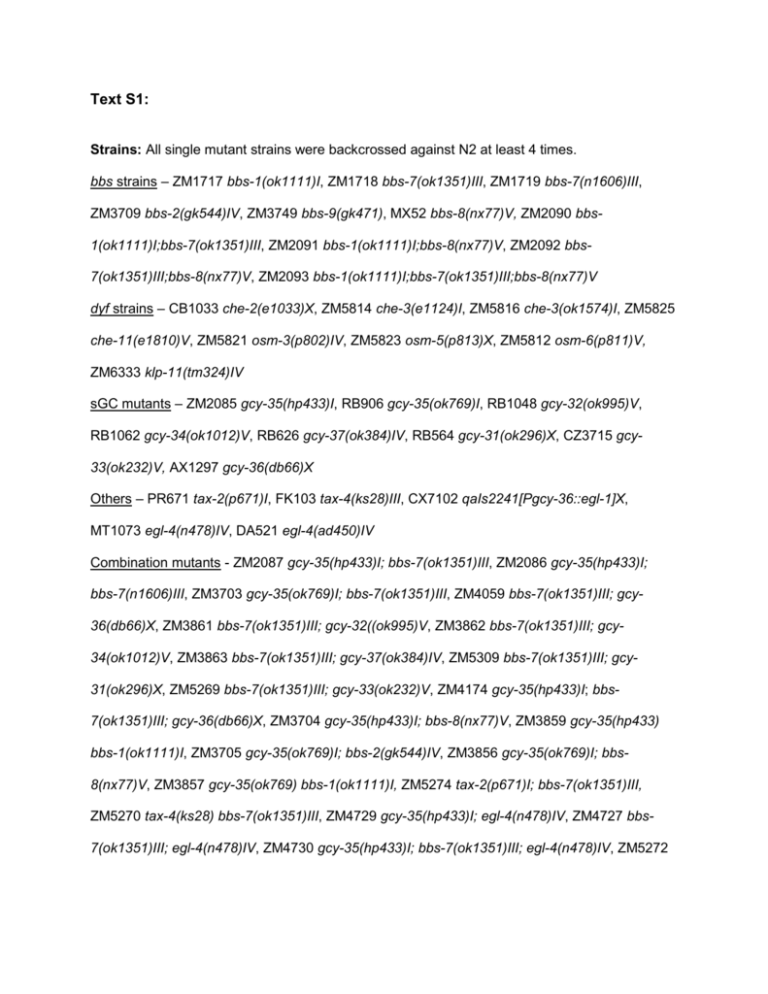
Text S1: Strains: All single mutant strains were backcrossed against N2 at least 4 times. bbs strains – ZM1717 bbs-1(ok1111)I, ZM1718 bbs-7(ok1351)III, ZM1719 bbs-7(n1606)III, ZM3709 bbs-2(gk544)IV, ZM3749 bbs-9(gk471), MX52 bbs-8(nx77)V, ZM2090 bbs1(ok1111)I;bbs-7(ok1351)III, ZM2091 bbs-1(ok1111)I;bbs-8(nx77)V, ZM2092 bbs7(ok1351)III;bbs-8(nx77)V, ZM2093 bbs-1(ok1111)I;bbs-7(ok1351)III;bbs-8(nx77)V dyf strains – CB1033 che-2(e1033)X, ZM5814 che-3(e1124)I, ZM5816 che-3(ok1574)I, ZM5825 che-11(e1810)V, ZM5821 osm-3(p802)IV, ZM5823 osm-5(p813)X, ZM5812 osm-6(p811)V, ZM6333 klp-11(tm324)IV sGC mutants – ZM2085 gcy-35(hp433)I, RB906 gcy-35(ok769)I, RB1048 gcy-32(ok995)V, RB1062 gcy-34(ok1012)V, RB626 gcy-37(ok384)IV, RB564 gcy-31(ok296)X, CZ3715 gcy33(ok232)V, AX1297 gcy-36(db66)X Others – PR671 tax-2(p671)I, FK103 tax-4(ks28)III, CX7102 qaIs2241[Pgcy-36::egl-1]X, MT1073 egl-4(n478)IV, DA521 egl-4(ad450)IV Combination mutants - ZM2087 gcy-35(hp433)I; bbs-7(ok1351)III, ZM2086 gcy-35(hp433)I; bbs-7(n1606)III, ZM3703 gcy-35(ok769)I; bbs-7(ok1351)III, ZM4059 bbs-7(ok1351)III; gcy36(db66)X, ZM3861 bbs-7(ok1351)III; gcy-32((ok995)V, ZM3862 bbs-7(ok1351)III; gcy34(ok1012)V, ZM3863 bbs-7(ok1351)III; gcy-37(ok384)IV, ZM5309 bbs-7(ok1351)III; gcy31(ok296)X, ZM5269 bbs-7(ok1351)III; gcy-33(ok232)V, ZM4174 gcy-35(hp433)I; bbs7(ok1351)III; gcy-36(db66)X, ZM3704 gcy-35(hp433)I; bbs-8(nx77)V, ZM3859 gcy-35(hp433) bbs-1(ok1111)I, ZM3705 gcy-35(ok769)I; bbs-2(gk544)IV, ZM3856 gcy-35(ok769)I; bbs8(nx77)V, ZM3857 gcy-35(ok769) bbs-1(ok1111)I, ZM5274 tax-2(p671)I; bbs-7(ok1351)III, ZM5270 tax-4(ks28) bbs-7(ok1351)III, ZM4729 gcy-35(hp433)I; egl-4(n478)IV, ZM4727 bbs7(ok1351)III; egl-4(n478)IV, ZM4730 gcy-35(hp433)I; bbs-7(ok1351)III; egl-4(n478)IV, ZM5272 gcy-35(hp433)I; egl-4(ad450)IV, ZM5273 bbs-7(ok1351)III; egl-4(ad450)IV, ZM5271 gcy35(hp433)I; bbs-7(ok1351)III; egl-4(ad450)IV rGC combination mutants – ZM5725 gcy-1(tm2669)II;bbs-7(ok1351)III, ZM5726 gcy4(tm1653)II;bbs-7(ok1351)III, ZM5613 gcy-5(ok930)II;bbs-7(ok1351)III, ZM5727 bbs7(ok1351)III;gcy-6(tm1449)V, ZM5728 bbs-7(ok1351)III;gcy-7(tm901)V, ZM5465 bbs7(ok1351)III;gcy-8(oy44)IV, ZM5729 bbs-7(ok1351)III;gcy-9(tm2816)X, ZM5614 bbs7(ok1351)III;gcy-16(ok2538)V, ZM5466 bbs-7(ok1351)III;gcy-18(nj38)IV, ZM5730 bbs7(ok1351)III;gcy-22(tm2364)V, ZM5612 bbs-7(ok1351)III;gcy-23(ok797)IV, ZM5731 bbs7(ok1351)III;gcy-5(tm4300)V, ZM5732 bbs-7(ok1351)III;gcy-28(tm2411)I, ZM5615 bbs7(ok1351)III; gyc-8(oy44) gcy-18(nj38), ZM5733 bbs-7(ok1351)III; gcy-23(ok797) gyc-8(oy44) gcy-18(nj38), ZM5467 bbs-7(ok1351)III;odr-10(n1936)X, ZM4928 bbs-7(ok1351)III;daf11(m47)V, ZM5616 bbs-7(ok1351)III;daf-11(ks67)V, ZM5991 gcy-35(hp433)I;gcy-23(ok797)IV, ZM5992 gcy-35(hp433)I;gcy-23(ok797)IV;bbs-7(ok1351)III, ZM5993 gcy-35(hp433)I;gcy4(tm1653)II, ZM5994 gcy-35(hp433)I;gcy-4(tm1653)II;bbs-7(ok1351)III, ZM5996 gcy35(hp433)I;bbs-7(ok1351)III;gcy-25(tm4300)IV, ZM5997 gcy-28(tm2411) gcy-35(hp433)I; bbs7(ok1351)III, ZM5998 gcy-35(hp433)I;gcy-16(ok2538)V, ZM5999 gcy-35(hp433)I;bbs7(ok1351)III;gcy-16(ok2538)V, ZM6000 gcy-35(hp433)I;gcy-7(tm901)V, ZM6001 gcy35(hp433)I;bbs-7(ok1351)III;gcy-7(tm901)V dyf combination mutants – ZM5817 gcy-35(hp433)I;che-2(e1033)X, ZM5815 gcy-35(hp433) che-3(e1124)I, ZM5818 gcy-35(hp433) che-3(ok1574)I, ZM5826 gcy-35(hp433)I;che11(e1810)V, ZM5822 gcy-35(hp433)I;osm-3(p802)IV, ZM5824 gcy-35(hp433)I;osm-5(p813)X, ZM5813 gcy-35(hp433)I;osm-6(p811)V, ZM6334 gcy-35(hp433)I;klp-11(tm324)IV Transgenic Strains: Extrachromosomal arrays (hpEx lines) were generated by co-injecting various DNA constructs at ~10-20ng/ul with co-injection markers at 10ng/ul. The following are the strains and genotypes of the hpEx lines used in this study: Rescuing lines for bbs animals – ZM(1990, 1992) bbs-7(n1606)III; hpEx(463,465)[bbs7(wt)+Podr-1::GFP]), ZM2001 bbs-7(ok1351)III; hpEx472[bbs-7(wt)+Podr-1::GFP], ZM(24602463) bbs-8(nx77)V; hpEx(636-639)[bbs-8(wt)+Podr-1::GFP], ZM(2464-2470) bbs-1(ok1111)I; hpEx(640-646)[bbs-1(wt)+Podr-1::GFP], ZM(4007-4008) bbs-2(gk544)IV; hpEx(15511552)[pJH1516+Podr-1::GFP], ZM2459 gcy-35(hp433)I; bbs-7(n1606)III; hpEx635[bbs-7+Podr1::GFP], ZM5522 bbs-2(gk544) hpIs213(Pgcy-36::GFP::BBS-2)IV Rescuing lines for gcy-35(hp433)I;bbs-7(ok1351)III animals – ZM3390 gcy-35(hp433)I; bbs7(ok1351)III; hpEx1251[WRM0641cB09+Podr-1], ZM3391 gcy-35(hp433)I; bbs-7(ok1351)III; hpEx1252[WRM063aC10+Podr-1]), ZM(3393-3394) gcy-35(hp433)I;bbs-7(ok1351)III; hpEx(1256-1257)[WRM0613cC03+Podr-1], ZM3395 gcy-35(hp433)I; bbs-7(ok1351)III; hpEx1262[WRM0641cB09/MluI+Podr-1]). Cell-specific rescue lines for gcy-35(hp433)I;bbs-7(ok1351)III animals – ZM(3629-3632) gcy35(hp433)I;bbs-7(ok1351)III; hpEx(1360-1363)[Pgcy-36::GCY-35(non-isoprenylated)+Podr-1], ZM(3633-3635) gcy-35(hp433)I;bbs-7(ok1351)III; hpEx1364-1366[Plad-2::GCY35(isoprenylated)+Podr-1], ZM(3747-3748) gcy-35(hp433)I; bbs-7(ok1351)III; hpEx(13671368)[pJH1374+Podr-1], ZM(4062-4063) gcy-35(hp433)I; bbs-7(ok1351)III; hpEx(15681569)[pJH1493+Podr-1] Transgenic lines to examine the subcellular localisation of GCY-35 and GCY-36 – ZM4064 gcy36(db66)X; hpEx1570[Pgcy-36::GFP::GCY-36+Podr-1::GFP], ZM4065 bbs-7(ok1351)III; gcy36(db66)X; hpEx1571[Pgcy-36::GFP::GCY-36+ Podr-1::GFP], ZM5540 gcy-35(hp433)I; hpEx2312[pJH1867+Podr-1::GFP], ZM(5538-5539) gcy-35(hp433)I; bbs-7(ok1351)III; hpEx(2310-2311)[pJH1867+Podr-1::GFP], ZM5529 hpIs215[Pgcy-36::GFP::GCY-36](X), ZM5530 bbs-7(ok1351)III; hpIs215(X) EGL-4 gain of function expression lines – ZM(5534-5536) gcy-35(hp433)I; bbs-7(ok1351)III; hpEx(2306-2308)[Pgcy-36::egl-4(ad450)+Podr-1::GFP], ZM5537 N2; hpEx2309[Pgcy-36::egl4(ad450)+Podr-1::GFP] Molecular biology: Genomic rescues of bbs-1, bbs-7, and bbs-8 used PCR products of each respective genomic sequence, including 2080, 1262, and 531bp of 5’ upstream sequence respectively. Rescue of bbs-2 animals used bbs-2 cDNA (F20D12.3 ordered from OpenBiosystems, catalog # OCE1182-7243861) driven by an F25B3.3 (rgef-1) promoter sequence of 3444bp. gcy-35 rescues were carried out with clones Pgcy-36::GCY-35 (nonisoprenylated) [1] and Plad-2::GCY-35 (isoprenylated) [2] courtesy of Dr. Cornelia Bargmann. Promoters for these two plasmids were 1095bp and 4013bp respectively and exchanged using FseI and AscI sites to generate pJH1374 (Pgcy-36::GCY-35 isoprenylated) and pJH1390 (Plad2::GCY-35 non-isoprenylated). pJH1493 (Pflp-8::GCY-35 isoprenylated) was created by insertion of a FseI/AscI tagged, 2.3kb sequence 5’ of the flp-8 start site PCR’d from N2 genomic DNA (p3548: ATTggccggccTCAGAAACCCCGATTCAAAC, p3549: gtcGGCGCGCCTTTCTACTTG AAAAGTGTGGAC). Subcellular localisation of GCY-36 was analysed using a vector of Pgcy-36::GFP::GCY36 [3] courtesy of Dr. Mario de Bono that included 2021bp of upstream promoter sequence. pJH1867 (Pgcy-36::GFP::GCY-35) was constructed by replacing GCY-36 in the above construct with a subcloned GCY-35 from pJH1374 using p3639 SfoI-tagged (ATTGGCGCCacaATGTTCGGCT GGATTCACG) and p3640 HpaI-tagged (GTCgttaacttaAGAAATTGTGCAAGTCG) primers. Phenotype analyses of C. elegans wild-type and mutant animals DiI uptake - DiI assays were performed on mixed populations of strains as previously described [4]. C. elegans were washed twice in M9 buffer and resuspended in 400ul M9 with 2ng of DiI. Samples were incubated 2-3 hours before washing three times with M9 buffer. Worms were allowed to recover overnight before scoring for uptake phenotypes. Uptake was scored as positive in an animal if DiI staining was observed in amphid, phasmid or both sets of neurons on a minimum of 20 animals per strain. Fisher’s Exact test was used to compare rescues between wildt-type and bbs strains as well as between bbs and bbs rescue strains. Body size assessment - Three gravid adult animals per strain were allowed to lay eggs for 2430 hours before being removed from each plate. F1 animals were allowed to grow for an additional 48 hours. Approximately 20-40 L4 animals were measured for each strain. L4 animals were allowed to grow 16-22 hours before measurement for adult data. Some populations were measured again 48 hours later to complete growth curve analysis. For body size measurement, images were captured on a Zeiss Stemi SVII dissecting microscope at 50X magnification using a Zeiss Axiocam digital camera at a 1388x1040 resolution. These images were then analysed using Axiovision software to measure length (cubic splines) and/or width of individual animals. Statistical analyses were completed using ANOVA with the Tukey post-hoc test to compare between multiple groups; *** p<0.001 ** p<0.01, * p<0.05, and ns p≥0.05. Statistical significance is only noted for a body size difference of >3.5% of wild-type length. Data on all graphs represent mean ± standard deviation relative to wild-type body length. Developmental timing - The developmental timing assay was modified from [5]. Briefly, eggs were collected from gravid adults and hatched in 7.5mL M9 for 20-22 hours. Synchronized L1 larvae were centrifuged again and animals were deposited onto multiple OP50-seeded NGM plates at about 50-80 animals per plate. Animals were grown at 20ºC for ~42 hours before being assessed for developmental staging based on described anatomical landmarks for L4 larvae at 30-minute intervals. Each plate was analysed as a single population, with at least 10 replicate plates per strain. The ANOVA with Tukey post-hoc test was used for statistical analyses; *** p<0.001 ** p<0.01, * p<0.05, and ns p≥0.05 in comparing between strains. Roaming assay – Animals at the L4 stage were transferred individually to the centre of 35mm seeded plates. Individual animals were removed 18 hours later and their tracking pattern was analysed using a 5mm x 5mm grid. Inclusion criteria for grids were any bacterial squares that the animals had traversed in the given time. For each strain a minimum of 25 animals was examined. Statistical analyses were completed using the Kruskal-Wallis non-parametric test with Dunn’s post-hoc to compare between multiple groups; *** p<0.001 ** p<0.01, * p<0.05, and ns p≥0.05. Data on all graphs represent mean ± standard error of mean (SEM) for squares traversed. Fluorescence Microscopy Animals were bleach synchronized and examined at various stages including at L3, L4 and 1220 hours later. Animals were immobilised on 2-5% dry agarose imaging pads, with 2ul of M9 solution. All images were acquired on a Zeiss Axioskop 2 fluorescent microscope at 63x magnification equipped with a CCD camera. Supplemental References 1. Gray JM, Karow DS, Lu H, Chang AJ, Chang JS, et al. (2004) Oxygen sensation and social feeding mediated by a C. elegans guanylate cyclase homologue. Nature 430: 317-322. 2. Chang AJ, Chronis N, Karow DS, Marletta MA, Bargmann CI (2006) A distributed chemosensory circuit for oxygen preference in C. elegans. PLoS Biol 4: e274. 3. Cheung BH, Arellano-Carbajal F, Rybicki I, de Bono M (2004) Soluble guanylate cyclases act in neurons exposed to the body fluid to promote C. elegans aggregation behavior. Curr Biol 14: 1105-1111. 4. Uchida O, Nakano H, Koga M, Ohshima Y (2003) The C. elegans che-1 gene encodes a zinc finger transcription factor required for specification of the ASE chemosensory neurons. Development 130: 1215-1224. 5. Sulston JE, A. HJ (1988) Methods. The Nematode Caenorhabditis elegans. New York: Cold Spring Harbor Laboratory Press. pp. 587-606.


