USMLE Step 1 Web Prep — Major Histocompatability Complex
advertisement
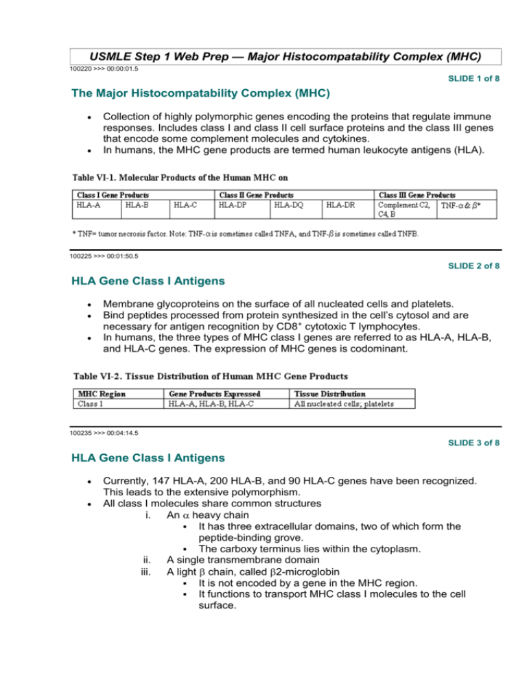
USMLE Step 1 Web Prep — Major Histocompatability Complex (MHC) 100220 >>> 00:00:01.5 SLIDE 1 of 8 The Major Histocompatability Complex (MHC) Collection of highly polymorphic genes encoding the proteins that regulate immune responses. Includes class I and class II cell surface proteins and the class III genes that encode some complement molecules and cytokines. In humans, the MHC gene products are termed human leukocyte antigens (HLA). 100225 >>> 00:01:50.5 SLIDE 2 of 8 HLA Gene Class I Antigens Membrane glycoproteins on the surface of all nucleated cells and platelets. Bind peptides processed from protein synthesized in the cell’s cytosol and are necessary for antigen recognition by CD8+ cytotoxic T lymphocytes. In humans, the three types of MHC class I genes are referred to as HLA-A, HLA-B, and HLA-C genes. The expression of MHC genes is codominant. 100235 >>> 00:04:14.5 SLIDE 3 of 8 HLA Gene Class I Antigens Currently, 147 HLA-A, 200 HLA-B, and 90 HLA-C genes have been recognized. This leads to the extensive polymorphism. All class I molecules share common structures i. An heavy chain It has three extracellular domains, two of which form the peptide-binding grove. The carboxy terminus lies within the cytoplasm. ii. A single transmembrane domain iii. A light chain, called 2-microglobin It is not encoded by a gene in the MHC region. It functions to transport MHC class I molecules to the cell surface. 100240 >>> 00:06:04.5 SLIDE 4 of 8 The MHC I Endogenous Pathway Endogenously produced proteins (eg. from viruses, intracellular bacteria, transformed tumor cells) degraded by proteases Peptide products transported to TAP complex, then to the endoplasmic reticulum (ER) In ER, peptides bind to HLA (MHC) class I proteins Cytotoxic CD8+ T lymphocytes recognize peptides that are associated with class I protein (MHC restriction). 100245 >>> 00:07:59.5 SLIDE 5 of 8 HLA Gene Class II Antigens HLA class II proteins are expressed on antigen-presenting cells. Bind exogenous peptide epitopes that have been endocytosed and processed Necessary for antigen recognition by CD4+ helper T lymphocytes. Class II genes code for cell-surface glycoproteins with two polypeptide components called and . In humans, the class II genes include HLA-DR, HLA-DQ, and HLADP. As with the class I system, the class II genes are highly polymorphic. They are also codominantly expressed. 100250 >>> 00:10:09.5 SLIDE 6 of 8 In a Nutshell: Class II Molecules Internalized and digested exogenous antigens; the resultant fragments complex with class II molecules to be presented on the cell surface. Both and chains with two extracellular domains. Two transmembrane domains. Recognized by CD4+ helper T cells. 100255 >>> 00:11:33.5 SLIDE 7 of 8 MHC II APCs endocytose and process foreign protein into small (about 10–20 amino acid) peptides. The MHC class II and chains synthesized with invariant chain. Role of invariant chain is to block access to peptide-binding groove until released and degraded once the vesicle containing the MHC class II molecule fuses with endocytic vacuole. MHC class II molecules reacts with degraded peptides. CD4+ helper T cells have antigen receptors that recognize only peptides bound to class II molecules (MHC restriction). 100260 >>> 00:12:46.5 SLIDE 8 of 8 A 21-year-old man presents with cough, fever, and hemoptysis. Blood tests show significantly elevated BUN and creatinine. Immunofluorescent microscopy reveals a diffuse linear pattern of fluorescence along the basement membranes of alveolar septa and glomerular capillaries. Which type of hypersensitivity is associated with this disease? (A) Type I (B) Type II (C) Type III (D) Type IV Explanations: The correct answer is B. This patient has a USMLE-favorite disease: Goodpasture's syndrome, which affects both the renal and pulmonary systems. In the kidney, it causes a rapidly progressive glomerulonephritis caused by antibodies directed against a collagen component of the glomerular basement membrane (anti-GBM antibody-a classic clue to this diagnosis). These antibodies create a linear pattern on immunofluorescence. Note that they are also active against the basement membrane of respiratory alveoli, accounting for the pulmonary component of the disease. Autoimmune reactions such as those found in Goodpasture's, certain drug allergies, blood transfusion reactions, and hemolytic disease of the newborn are classified as Type II hypersensitivities (antibody-mediated cytotoxicity). IgG or IgM antibody reacts with membrane-associated antigen on the surface of cells, causing activation of the complement cascade and, ultimately, cell destruction. Type I (choice A) (immediate, atopic, or anaphylactic) reactions require an initial (sensitizing) exposure to an antigen. Upon re-exposure to the antigen, cross-linking of IgE receptors occurs on the surface of basophils and mast cells. The mast cells then release a variety of mediators, including histamine. Clinical syndromes include asthma, atopic dermatitis, eczema, and allergic rhinitis. Hives ("wheal and flare") are characteristic of Type I hypersensitivity. Type III (choice C) hypersensitivity (immune complex-mediated hypersensitivity) is caused by antibodies to foreign antigens. Immune complexes of IgG or IgM with the antigen activate complement, resulting in the generation of C3b, which promotes neutrophil adherence to blood vessel walls. The complexes also generate C3a and C5a (anaphylatoxins), which lead to inflammation and tissue destruction. The hallmark signs of Type III sickness occur 7-14 days after exposure to the offending antigen and include urticaria, angioedema, fever, chills, malaise, and glomerulonephritis. Clinical syndromes include serum sickness (e.g., penicillin, streptomycin, sulfonamide, phenylbutazone hypersensitivity) and the Arthus response. Immune complexes are also observed in lupus (SLE). Type III glomerulonephritis (e.g., poststreptococcal glomerulonephritis) is characterized by a "lumpy bumpy" appearance on immunofluorescence using tagged antibody specific for immunoglobulin or complement. Type IV (choice D) is also known as delayed-type hypersensitivity (DTH). Unlike the other types, which are mediated by antibody, DTH depends on TDTH cells that have been sensitized to a particular antigen. T cells react with antigen in association with MHC class I gene products and release lymphokines. Examples include tuberculin skin sensitivity and contact dermatitis (e.g., poison ivy rash).

