Staphylococcus intermedius in Canine Gingiva and Canine
advertisement
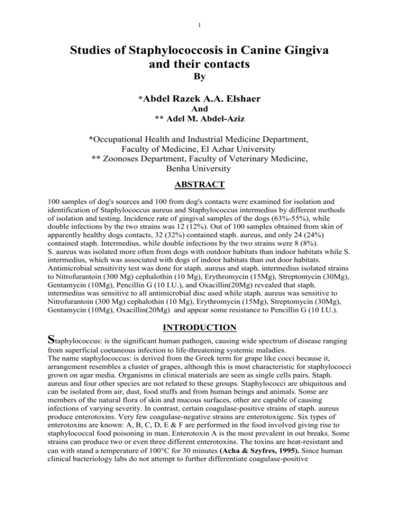
1 Studies of Staphylococcosis in Canine Gingiva and their contacts By *Abdel Razek A.A. Elshaer And ** Adel M. Abdel-Aziz *Occupational Health and Industrial Medicine Department, Faculty of Medicine, El Azhar University ** Zoonoses Department, Faculty of Veterinary Medicine, Benha University ABSTRACT 100 samples of dog's sources and 100 from dog's contacts were examined for isolation and identification of Staphylococcus aureus and Staphylococcus intermedius by different methods of isolation and testing. Incidence rate of gingival samples of the dogs (63%-55%), while double infections by the two strains was 12 (12%). Out of 100 samples obtained from skin of apparently healthy dogs contacts, 32 (32%) contained staph. aureus, and only 24 (24%) contained staph. Intermedius, while double infections by the two strains were 8 (8%). S. aureus was isolated more often from dogs with outdoor habitats than indoor habitats while S. intermedius, which was associated with dogs of indoor habitats than out door habitats. Antimicrobial sensitivity test was done for staph. aureus and staph. intermedius isolated strains to Nitrofurantoin (300 Mg) cephalothin (10 Mg), Erythromycin (15Mg), Streptomycin (30Mg), Gentamycin (10Mg), Pencillin G (10 I.U.), and Oxacillin(20Mg) revealed that staph. intermedius was sensitive to all antimicrobial disc used while staph. aureus was sensitive to Nitrofurantoin (300 Mg) cephalothin (10 Mg), Erythromycin (15Mg), Streptomycin (30Mg), Gentamycin (10Mg), Oxacillin(20Mg) and appear some resistance to Pencillin G (10 I.U.). INTRODUCTION Staphylococcus: is the significant human pathogen, causing wide spectrum of disease ranging from superficial coetaneous infection to life-threatening systemic maladies. The name staphylococcus: is derived from the Greek term for grape like cocci because it, arrangement resembles a cluster of grapes, although this is most characteristic for staphylococci grown on agar media. Organisms in clinical materials are seen as single cells pairs. Staph. aureus and four other species are not related to these groups. Staphylococci are ubiquitous and can be isolated from air, dust, food stuffs and from human beings and animals. Some are members of the natural flora of skin and mucous surfaces, other are capable of causing infections of varying severity. In contrast, certain coagulase-positive strains of staph. aureus produce enterotoxins. Very few coagulase-negative strains are enterotoxigenc. Six types of enterotoxins are known: A, B, C, D, E & F are performed in the food involved giving rise to staphylococcal food poisoning in man. Enterotoxin A is the most prevalent in out breaks. Some strains can produce two or even three different enterotoxins. The toxins are heat-resistant and can with stand a temperature of 100C for 30 minutes (Acha & Szyfres, 1995). Since human clinical bacteriology labs do not attempt to further differentiate coagulase-positive 2 staphylococcal species and since S. intermedius is clearly a skin pathogen in animals, this staphylococcal organism may be a human pathogen in canine-inflicted wounds. Some cases of S. intermedius associated with dog bite infections support this concept. S. intermedius was commonly found to be a gingival flora of the upper front teeth with S. aureus. However, S. aureus was more common among males, large dogs, and those of working breeds than was S. intermedius. S. intermedius and S. aureus were suggesting that the organisms may be mutually exclusive canine floras (Talan et al., 1989). To our knowledge, the isolates of S. Intermedius from dog bite wounds are the first such cases to be associated with human infection. From this previous study we isolated S. intermedius from an abscess in a human being (Adegoke, 1986). Another previous report which demonstrated enterotoxin-producing stains in dogs with S. intermedius suggested that this organism could have a pathogenic role in cases of human food poisoning and appears that S. intermedius may be a relatively common and potentially invasive pathogen of canine-inflicted human wounds (Fukuda et al., 1984). The current study was carried out to throw light on the following: 1-Prevalence of Staphylococcus aureus and Staphylococcus intermedius in gingival samples of dogs. 2- Prevalence of Staphylococcus aureus and Staphylococcus intermedius in gingival samples of man in contacts with these dogs. 3- Results of uses of antimicrobial sensitivity test to Staphylococcosis strains. MATERIALS AND METHODS In the present study 100 swabs were taken from gingival of dogs which were randomly collected from indoor and outdoor habits as well as100 swabs were obtained from skin of the palm of the hands of men occupational contacts with dogs examined. Again human samples were mainly taken from apparently normal persons and little from clinical ones. Sampling: Nasal and skin samples: Swabs for bacteriological examination were collected from the gingiva of dogs and skin of the palm of hands of human contacts. A sterile cotton swabs premoisted in sterile tubes containing nutrient broth were used for gingival swabs. The swabs then placed in test tubes containing 2ml nutrient broth. Human skin samples were swabbed with large swabs made by wrapping several layers of cotton on small artery forceps. The premoisted swabs were rubbed to-and-fro six times on the selected area the cotton wool pushed off into a cotton capped sterile test tube containing 10 ml nutrient broth. Collected swabs were carried to the laboratory with a minimum of delay in ice bag to be immediately examined. The interval between the collection of the swabs and the inoculation of primary plates was usually between 1.1/2 and 4 hr., and did not exceed 5hr. (Mahoudeau et al., 1997). Bacteriological Examination of the Collected Samples: Selective media for Staphylococci: Materials: Liquid medium. Nutrient broth (Gibco). Solid media. Barid-Parker's agar (oxoid). 3 Method: A loopful from Nutrient broth of pH (6.8-7) was streaked into plate of Baird-Parker's agar and examined after 48 hours incubation at 37C. Pathogenic colonies were black surrounded by halozone (Faint yellow in color). Identification of Zoonotic Staphylococci: Morphological Characters: I-Staining reaction: Films prepared from pure culture of isolated colonies were grape-like clusters with some single and paired cocci it is gram positive cocci; approximately 1mm in diameter non sporing and non capsulate. II-Biochemical Reactions: Biochemical identification of isolates: was made on the basis of the following tests according to (McFadden, 1980). S. intermedius, is clearly distinguishable from S. aureus by its microbiological and biochemical characteristics. S. intermedius was distinguished from S. aureus by the following characteristics: coagulation of rabbit plasma at 4h, hemolysis of sheep blood at 24h, and mannitol fermentation at 24h. A clear separation of the two species was apparent only with the acetoin (modified Voges-Proskauer) reaction and P galactosidase activity on the API Staph-Ident strip. Antimicrobial sensitivity test: More than one strain from the strains having the ability of cross infection between man and animals were used in the test against penicillin G 10 I.U. Penicillin resistant strains were tested again to different antibiotics to determines sensitivity and resistance. In vitro antimicrobial testing for the isolated and identified bacterial isolates were performed according to (Finegold and Martin, 1982). The procedure was as follows: A-Inoculation of plates: 1-Using a sterile loop, the top of four to five colonies were transferred to sterile test tube containing 5 ml of nutrient broth then incubated at 35C for about 18 hours. 2-A sterile cotton swab was dipped into the standard bacterial suspension and streaked onto the dried surface of nutrient agar with an even distribution of the inoculum. 3-The plates were covered and remained for 5 minutes for absorption of the excess moisture. 4-By using a sterile pointed forceps, the antibiotic discs were placed on the inoculated plate and pressed firmly. 5-The discs distributed evening in a manner that they were away from the edge of the petri-dish by 15 millimeter and the distance between the centers of two discs not less than 24 millimeter. B- Reading: After incubation at 35C for 24 hours, each plate was examined and the diameter of the complete inhibition zone (Clear Zone of inhibited growth around the discs) was measured by using reflected light and sliding caliper to the nearest millimeter. The diameter obtained zones of the bacterial isolates were interpreted by referring to the following table. Zone interpretive standards of National committee Clinical Laboratory Standards (NCCLS) (1984). Antimicrobic agent Disc potency Cephalothin Erythromycin Gentamycin Nitrofurantoin Oxacillin Pencillin G Streptomycin Results were recorded in table (4). 10Mg 15Mg 10Mg 300 Mg 20Mg 10 I.U 30Mg Inhibition zone 14 or less 14-17 12 or less 14 or less 13 or less 14 or less 14 or less Sensitive S S S S S S S 4 RESULTS Table (1): Staph. aureus and Staph. intermedius isolated from Gingival samples of examined Dogs: Staph. species Staph. aureus Staph. intermediu Total gingiva samples 100 100 Numbers of Percentage Positive of Positive Numbers of Percentage negative of negative 63 55 37 45 63% 55% Double infection 37% 45% 12 Percentage of Double infection 12% Table (2): Staph. aureus and Staph. intermedius isolated from skin of Dogs attendants: Total skin Staph. species samples Numbers of Positive Percentage of Numbers of Positive negative Percentage of negative Staph. aureus 100 100 Staph. intermediu 32 24 32% 24% 68% 76% 68 76 Double Percentage infection of double infections 8 8% Table (3): Differentiation between indoor and outdoor Staphylococcosis isolated from Gingival samples of examined Dogs: Types of Staphylococcus S. aureus S. intermedius Total positive samples 63 55 Indoor positive samples 28 15 Outdoor positive samples 35 40 Table (4) Summarized results of antimicrobial sensitivity test of Staphylococcosis strains: Antimicrobic agent Disc potency Inhibited zone for S.aureus Inhibited zone for S. intermedius Results for S.aureus Results for S. intermedius Cephalothin Erythromycin Gentamycin Nitrofurantoin Streptomycin Oxacillin Pencillin G 10 Mg 15 Mg 10 Mg 300 Mg 30 mg 20 Mg 10 IU 21 mm 19 mm 17 mm 20 mm 16 mm 15 mm 8 mm . 20 mm 16 mm 19 mm 22 mm 18 mm 17 mm 18 mm S S S S S S R S S S S S S S S = sensitive. R= resistance. 5 DISCUSSION In these investigation trials has been made to isolate staphylococci from man and dogs. Identification of the isolates was limited to S. intermedius and S. aureus isolates 100 Samples from different dog's source and 100 from human contacts were examined for isolation and identification of staph. aureus by using, Staining reaction, Coagulase tube test, hemolysis, Mannitol fermentation, modified Voges-Proskauer reaction, Catalase activity, Detection of Gelatin hydrolysis. Coagulase reactions were positive after incubation for 24 h for all S. intermedius and S. aureus isolates; the tests were positive after 4 h for 30% of the S. intermedius isolates, compared with 100% of the S. aureus Isolates. Delayed hemolysis, mannitol fermentation, and coagulation were generally characteristic of S. intermedius. However, a clear separation of the two species was possible only with the acetoin (modified Voges-Proskauer) reaction: 0% of the S. intermedius isolates were reactive, compared with 100% of S. aureus isolates. The result displayed in table (1) revealed that out of 100 gingival samples collected from apparently healthy dogs, staph. aureus was isolated from 63 (63%). This finding explains the importance of dogs as carriers for staph. aureus in their oral cavity. Incidence rate of carrier-states in examined dogs was 63%; a result coincides with that reported by (Cavaclcanti et al., 2005). Also the result displayed in the same table revealed that out of 100 gingival samples collected from apparently healthy dogs, staph. intermedius was isolated from 55 (55%). While double infected dogs by the two species are 12 (12%). This finding explains the importance of dogs as carriers for staph. intermedius in their oral cavity. Incidence rate of carrier-states in examined dogs was 55%, a result coincides with that reported by (Zdovc et al., 2004). The PH tolerance provides staphylococci a competitive advantage among the flora of the oral cavity the difference in incidence rates may be attributed to the hygienic state of dogs housing, level of environmental hygiene, care and proper management to the dogs. Table (2) shows that out of 100 samples obtained from skin of apparently healthy dog's contacts only 32 (32%) contained staph. aureus, While 24 (24%) contained staph. intermedius. While double infected dogs contacts by the two species are 8 (8%). This finding coincides to a certain extent with that reported by (Cizek and Bardon, 2006). Staphylococci have competitive advantage in hair follicles, sebaceous and sweat glands because they produce lipases and esterases and have the ability to grow under partial anaerobic conditions. It is note worthy that the skin carrier serves as a disperser of staphylococcus, largely as a result of the ability of organisms to transfer to the skin, clothing and bedding of the carrier himself. The result displayed in table (3) revealed that from100 gingival samples collected from apparently healthy dogs, staph. aureus was isolated in about 28, 35 indoor and outdoor respectively while in staph. intermedius about 15, 40 respectively. S. aureus was found significantly more often among dogs of indoor habitats than S. intermedius. This phenomenon may in part be explained by differences in the degrees of exposure of these dogs to other animals and human beings, as this has been shown to affect the carriage rates of these staphylococcal species among dogs. The difference in incidence rates may be attributed to the hygienic state of animal housing, level of environmental hygiene, care and proper management to the animals. The antibiotic susceptibilities of S. intermedius and S. aureus isolates are presented in Table (4) S. intermedius was susceptible to a wide range of antibiotics without exception, to which 100% of the isolates were sensitive. S. aureus had an antibiotic susceptibility pattern similar to that of S. intermedius except 100% of the S. aureus isolates were resistant to penicillin. There was no relationship between prior exposure to antibiotics and antibiotic resistance for dogs with S. intermedius or S. aureus. S. intermedius is more often susceptible to penicillin, and this may explain the findings of previous studies which have revealed a high incidence of penicillin-susceptible coagulase-positive staphylococci from canine specimens.This finding coincides to a certain extent with that reported by (Gerstadt et al., 1999) and (Hoekstra and Paulton, 2002). 6 REFERENCES Acha P. N. and Szyfres B., (1995) Zoonoses and Communicable Diseases Common to Man and Animals. Pan American Health Organization Washington D.C.20037, U.S.A Adegoke, G. O., (1986). Characteristics of staphylococci isolated from man, poultry, and some other animals. J. Appl. Bacteriol.60:97-102. Cavalcanti S. M., Franca E.R., Cabral C., Vilela M.A. ,Montenegro F., Menezes D. and Medeiros A.C. (2005). Prevalence of Staphylococcus aureus introduced into intensive care units of a University Hospital. Braz J. infect. Dis. Feb; 9(1):56-63. Cizek A. and Bardon J., (2006) Zoonotic importance of selected species of Gram-positive bacteria. Klin. Mikrobiol.infekc.lek.12(1):10-12. Finegold S.M and Martin, W.J.(1982). Baily and Scott’s Diagnostic Microbiology.6th end. the C.V. Mosby Co./st. Lewis, Toranto, London Fukuda, S., Tokuno H., Ogawa H., Sasaki M., Kawano J., Shimizu A., and Kimura A. (1984). Enterotoxigenicity of Staphylococcus intermedius strains isolated from dogs. Zentralbl. Bakteriol. Mikrobiol. Hyg. A 258:360-367. Gerstadt, K., J. S. Daly, M. Mitchell, M. Wessolossky, and S. H. Cheeseman. (1999). Methicillin-resistant Staphylococcus intermedius pneumonia following coronary artery bypass grafting. Clin. Infect. Dis. 29:218219. Hoekstra K. A. and Paulton R.J., (2002) Clinical prevalence and antimicrobial susceptibility of Staphylococcus aureus and Staph. intermedius in dog's .J Appl. Microbiol; 93(3):406-13. MacFaddin J.F., (1980). Biochemical tests for identification of medical bacteria. The Williams and Wilkins Company Balatmore, U.S.A. Mahoudeau, I., Delabranche X., Prevost G., Monteil H., and Piemont Y., (1997). Frequency of isolation of Staphylococcus intermedius from humans. J. Clin. Microbiol. 35:2153-2154. National Committee for Clinical Laboratory Standards., (1984). Performance standards for antimicrobial disk susceptibility tests, M2-A3. National Committee for Clinical Laboratory Standards, Villanova, Pa. Talan D. A., Staatz D., Staatz A., and Overturf G. D., (1989). Staphylococcus intermedius: clinical presentation of a new human dog bite pathogen. Ann. Emerg. Med. 18:410-413. Zdovc I., ocepek M., Pirs T., Krt B., and Pinter L., (2004). Microbiological features of Staphylococcus schleiferi subsp. coagulans, isolated from dogs and possible misidentification with other canine coagulasepositive staphylococci. J. Vet. Med. B. infect. Dis. Vet. public Health Dec; 51(10):449-54.
