agctt atg ctg ggg ccc tgc atg ctg ctg ctg ctg ctg ctg ctg ggc ctg agg cta
advertisement
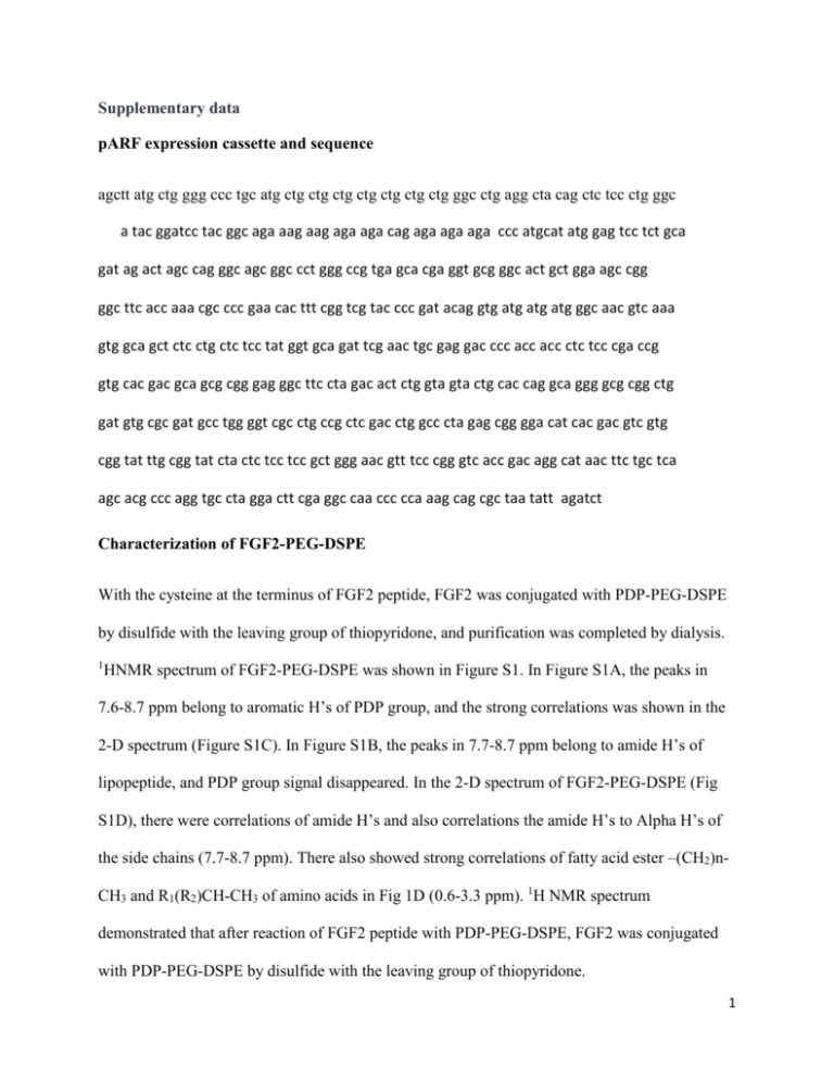
Supplementary data pARF expression cassette and sequence agctt atg ctg ggg ccc tgc atg ctg ctg ctg ctg ctg ctg ctg ggc ctg agg cta cag ctc tcc ctg ggc a tac ggatcc tac ggc aga aag aag aga aga cag aga aga aga ccc atgcat atg gag tcc tct gca gat ag act agc cag ggc agc ggc cct ggg ccg tga gca cga ggt gcg ggc act gct gga agc cgg ggc ttc acc aaa cgc ccc gaa cac ttt cgg tcg tac ccc gat acag gtg atg atg atg ggc aac gtc aaa gtg gca gct ctc ctg ctc tcc tat ggt gca gat tcg aac tgc gag gac ccc acc acc ctc tcc cga ccg gtg cac gac gca gcg cgg gag ggc ttc cta gac act ctg gta gta ctg cac cag gca ggg gcg cgg ctg gat gtg cgc gat gcc tgg ggt cgc ctg ccg ctc gac ctg gcc cta gag cgg gga cat cac gac gtc gtg cgg tat ttg cgg tat cta ctc tcc tcc gct ggg aac gtt tcc cgg gtc acc gac agg cat aac ttc tgc tca agc acg ccc agg tgc cta gga ctt cga ggc caa ccc cca aag cag cgc taa tatt agatct Characterization of FGF2-PEG-DSPE With the cysteine at the terminus of FGF2 peptide, FGF2 was conjugated with PDP-PEG-DSPE by disulfide with the leaving group of thiopyridone, and purification was completed by dialysis. 1 HNMR spectrum of FGF2-PEG-DSPE was shown in Figure S1. In Figure S1A, the peaks in 7.6-8.7 ppm belong to aromatic H’s of PDP group, and the strong correlations was shown in the 2-D spectrum (Figure S1C). In Figure S1B, the peaks in 7.7-8.7 ppm belong to amide H’s of lipopeptide, and PDP group signal disappeared. In the 2-D spectrum of FGF2-PEG-DSPE (Fig S1D), there were correlations of amide H’s and also correlations the amide H’s to Alpha H’s of the side chains (7.7-8.7 ppm). There also showed strong correlations of fatty acid ester –(CH2)nCH3 and R1(R2)CH-CH3 of amino acids in Fig 1D (0.6-3.3 ppm). 1H NMR spectrum demonstrated that after reaction of FGF2 peptide with PDP-PEG-DSPE, FGF2 was conjugated with PDP-PEG-DSPE by disulfide with the leaving group of thiopyridone. 1 Stable serum ability of GLL It is important to be serum stable as a gene delivery system for in vivo test. The transfection of pLUC by GLL on U87-MG cells growth demonstrated that the luciferase expression of the plasmid was higher when transfected in the media with serum than that of media without serum (Fig S2), which indicated that the delivery system GLL was stable in the media with serum. The higher gene expression was contributed by two factors. One is that the DNA-GLL complex was stable in the media with serum, and the cellular uptake of the positively charged lipoplex happened fast (0-2h, data not shown). The other factor is that the presence of serum maintains the cell in higher viability which favors gene expression. This phenomenon has been reported by [43]. 2 Supplementary figure legend: Fig S1 1H NMR spectrum (500 MHz) of PDP-PEG2000-DSPE and FGF2-PEG2000-DSPE a: PDP-PEG-DSPE, 1-Dimension spectrum; b: FGF2-PEG-DSPE, 1-Dimension spectrum; c: PDP-PEG-DSPE, 2-Dimension spectrum; d: FGF2-PEG-DSPE, 2-Dimension spectrum. Fig S2 Transfection of pLUC-GLL on U87-MG cells in DMEM media with (serum +) or without serum (serum-). N=3. P<0.1, student t-test, paired. 3

