Liver Tumors
advertisement
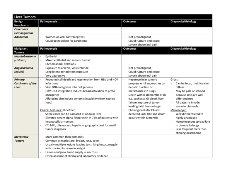
Liver Tumors Benign Neoplasms Cavernous Hemangiomas Adenomas Pathogenesis Outcomes - - Malignant Tumors Hepatoblastoma (children) Pathogenesis Angiosarcoma (adults) Primary Carcinoma of the Liver - Women on oral contraceptives Could be mistaken for carcinoma Epithelial Mixed epithelial and mesenchymal Chromosomal deletions Exposure to arsenic, vinyl chloride Long latent period from exposure Very aggressive Repeated cell death and regeneration from HBV and HCV infections Viral DNA integrates into cell genome HBV DNA integration induces broad activation of proto oncogenes Alfatoxins also induce genomic instability (from spoiled food) Clinical Features: Ill-defined - Some cases can be palpated as nodular liver - Elevated serum alpha-fetoprotein in 75% of patients with hepatocellular tumors - CT, MRI, ultrasound, hepatic angiography best for small tumor diagnosis Metastatic Tumors - More common than primaries Common primaries are: breast, lung, colon Usually multiple lesions leading to striking hepatomegaly with marked increase in weight Lesions outgrow blood supply -> necrosis Often absence of clinical and laboratory evidence Not premalignant Could rupture and cause severe abdominal pain Outcomes - - - Not premalignant Could rupture and cause severe abdominal pain Hepatocellular tumors progress until encroaches on hepatic function or metastasizes to lungs. Death within 10 months of dx e.g. cachexia, GI bleed, liver failure, rupture of tumor leading fatal hemorrhage Cholangiocellular CA not detected until late and death occurs within 6 months Diagnosis/Histology Diagnosis/Histology Gross: - Can be focal, multifocal or diffuse - May be pale or stained because cells are well differentiated - All patterns invade vascular channels Microscopic: - Well differentiated to highly anaplastic - Hematogenous spread late in disease to lungs - Less frequent mets than cholangiocarcinoma Disorder Inheritance Defect in metabolism of bilirubin Clinical course Crigler-Naijar syndrome, type I Ar Absent UGT1 activity Fatal in neo-natal period Crigler-Naijar syndrome, type II AD, variable penetrance Decreased UGT1 activity Mild, occasional kernicterus Gilbert’s syndrome Ar Decreased UGT1 activity Innocuous Dubin-Johnson syndrome Ar Impaired biliary excretion of bilirubin Innocuous Rotor syndrome Ar Impaired biliary excretion of bilirubin Innocuous Unconjugated hyperbilirubinemia Conjugated hyperbilirubinemia Intrahepatic Biliary Tract Diseases Secondary Biliary Cirrhosis Primary Billiary Cirrhosis Primary Sclerosing Cholangitis Etiology Extrahepatic bile duct obstruction: biliary atresia, gallstones, stricture, carcinoma of pancreatic head Possibly autoimmune Unknown, possibly autoimmune; 50% to 70% associated with inflammatory bowel disease Sex predilection None Female to male, Female to male, 6:1 1:2 Symptoms and signs Pruritus, jaundice, malaise, dark urine, Same as secondary biliary light stools, hepatosplenomegaly cirrhosis; insidious onset Laboratory findings Conjugated hyperbilirubinemia, Same as secondary biliary Same as secondary biliary cirrhosis, plus elevated increased serum alkaline phosphatase, cirrhosis, plus elevated serum IgM serum IgM, hypergammaglobulinemia autoantibodies (especially M2 bile acids, cholesterol form of anti-mitochondrial antibody) Same as secondary biliary cirrhosis; insidious onset Important pathologic Prominent bile stasis in bile ducts, bile Dense lymphocytic infiltrate in Periductal portal tracts fibrosis, segmental stenosis findings before ductular proliferation with surrounding portal tracts with granulomatous of extrahepatic and intrahepatic bile ducts cirrhosis develops neutrophils, portal tract edema destruction of bile ducts

