Dr Alanna Easton`s Travelling Scholarship Report, April 2014
advertisement
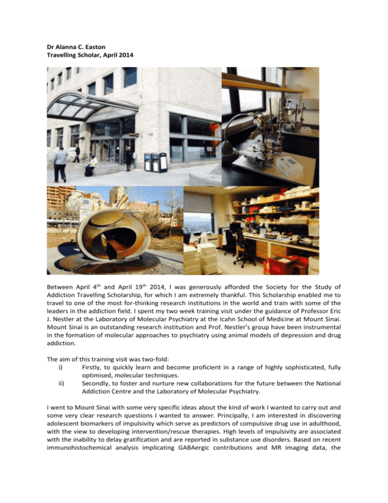
Dr Alanna C. Easton Travelling Scholar, April 2014 Between April 4th and April 19th 2014, I was generously afforded the Society for the Study of Addiction Travelling Scholarship, for which I am extremely thankful. This Scholarship enabled me to travel to one of the most for-thinking research institutions in the world and train with some of the leaders in the addiction field. I spent my two week training visit under the guidance of Professor Eric J. Nestler at the Laboratory of Molecular Psychiatry at the Icahn School of Medicine at Mount Sinai. Mount Sinai is an outstanding research institution and Prof. Nestler’s group have been instrumental in the formation of molecular approaches to psychiatry using animal models of depression and drug addiction. The aim of this training visit was two-fold: i) Firstly, to quickly learn and become proficient in a range of highly sophisticated, fully optimised, molecular techniques. ii) Secondly, to foster and nurture new collaborations for the future between the National Addiction Centre and the Laboratory of Molecular Psychiatry. I went to Mount Sinai with some very specific ideas about the kind of work I wanted to carry out and some very clear research questions I wanted to answer. Principally, I am interested in discovering adolescent biomarkers of impulsivity which serve as predictors of compulsive drug use in adulthood, with the view to developing intervention/rescue therapies. High levels of impulsivity are associated with the inability to delay gratification and are reported in substance use disorders. Based on recent immunohistochemical analysis implicating GABAergic contributions and MR imaging data, the central hypothesis of my work states that differential neuronal structure, and the specific pattern of development in the medium spiny neurons of the striatum will predict impulsivity. In agreement with my own research interests and given the relatively short amount of time I had at Mount Sinai, I focused my efforts on acquiring knowledge of the following experimental paradigms: Assessment of neuronal morphology, formation and reorganisation. This involved immunohistochemistry for medium spiny neuron visualisation using 100µm thick slices of brain tissue, looking in particular at the nucleus accumbens of mice. The method is used to detect antigens or proteins using antibodies which bind to these specific proteins in biological tissue. These antibodies are typically conjugated to an enzyme that can catalyse a colour-producing reaction, the fluorescence is then detected, in this case, by a confocal microscope. Using this technique, medium spiny neurons can be spatially located, selectively tagged and analysed for their spine shape and density, factors which are hypothesised to mediate impulsive behaviours. It is also possible to isolate different subtypes of medium spiny neurons, dopamine D1 expressing and dopamine D2 expressing, and assess the relative contribution of each subtype to the impulsivity phenotype. Differential growth factor activity specifically looking at neurotrophic factors to explain differential patterns of development of the medium spiny neurons of the striatum. The Western blotting technique is widely used to quantify protein changes in discreet brain regions, but delivers no information about the spatial location of such proteins. It uses gel electrophoresis to separate proteins based upon the length if their polypeptide chains. Once the proteins have been separated, they are stained with antibodies specific to the target protein, not dissimilar to the immunohistochemical technique used above, fluorescence is then detected and quantified using specially designed software. With the current research aims in mind, this technique will be useful in determining the expression levels of proteins including, BDNF, Trk B, NGF, NT-3 and NT-4 in discreet brain regions. These proteins are neurotrophic factors and could be instrumental in the progression of neuronal recruitment in the striatum. Chromatin modifications and protein interactions with DNA using quantitative ChIP and ChIP-seq. Chromatin immunoprecipitation (ChIP) is used to investigate the interaction between proteins and DNA in the cell. The technique aims to determine whether specific proteins are associated with specific genomic regions, such as transcription factors on promoter regions or other DNA binding sites. ChIP also aims to determine the specific location in the genome that various histone modifications are associated with, indicating the target of histone modifiers. In brief, the method uses fresh tissue and the protein and associated chromatin are temporarily bonded using a fixative. The tissue is then sheared while the protein-chromatin complex of interest retains its bond. The protein-chromatin fragments are immunoprecipitated, purified and released from one another. The chromatin sequences detected are therefore associated with the protein of interest in vivo. Histone modifications modulate the accessibility of DNA to transcription factors, which in turn control gene expression. Electrostatic interactions within the chromatin allow the DNA to ‘open’ and thus facilitate transcription. Alterations in gene expression are key and these changes will be relevant to my current research. In addition to learning the above mentioned techniques, I was able to observe some other paradigms and research projects the team were currently running, including: Optogenetic surgery and behaviour testing using channel rhodopsin to isolate and control neuronal excitation in discreet brain regions. Tissue extraction and preparation processes which will be prove to be very important for my work. Social defeat model of depression, an approach which has been shown to have excellent etiological, predictive, discriminative and face validity. I was also invited to weekly group and laboratory meetings to listen to members of the team discuss their current work. I also attended guest lectures and learnt about some of the exciting research that is currently taking place at other institutions and in association with Mount Sinai. This trip provided me with an excellent and extremely rare opportunity to work with some of the most dynamic people, and on some of the most stimulating and fascinating projects, in the addiction field. It also enabled me to meet personally with Prof. Nestler and hear his thoughts on the research programme I am currently developing. His input has been integral to my personal and professional development and I am eternally grateful of his offer of continued support. Prof. Nestler and his senior postdoctoral researchers, Dr Ja Wook Koo and Dr Mike Cahill, offered me state-of-the-art training in all aspects of the above techniques and generously offered me their protocols and their support and guidance going forward. I cannot thank them enough for this incredibly rewarding experience. The ultimate aim of the visit was for the techniques and approaches learnt to be implemented at the National Addiction Centre and performed in conjunction with my current research program. I hope to supervise and provide training to those colleagues, research staff and students which may wish to undertake similar tasks in the department. The visit also served to forge a new collaboration between Kings College London and Mount Sinai for future research at both establishments. I feel that this has been achieved with tremendous success and I am eternally grateful to the SSA executive for seeing the merit in my visit and providing me with the funds to be able to follow it through. The experience renewed my motivation and enthusiasm for my research and I look forward to the exciting collaborations that will follow in the future. Yours sincerely, Dr Alanna C. Easton National Addictions Centre, Institute of Psychiatry, Kings College London alanna.c.easton@kcl.ac.uk
