File - Science with Shust
advertisement
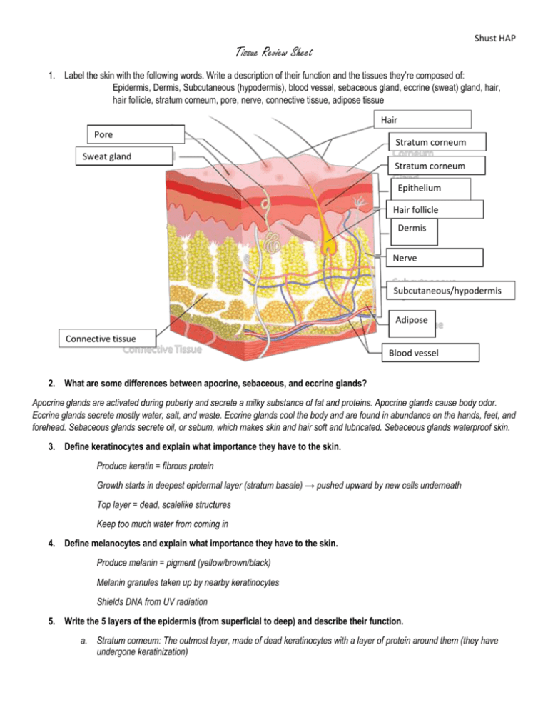
Shust HAP Tissue Review Sheet 1. Label the skin with the following words. Write a description of their function and the tissues they’re composed of: Epidermis, Dermis, Subcutaneous (hypodermis), blood vessel, sebaceous gland, eccrine (sweat) gland, hair, hair follicle, stratum corneum, pore, nerve, connective tissue, adipose tissue Hair Pore Connective tissue Sweat gland Stratum corneum Stratum corneum Connective tissue Stratum corneum Stratum corneum Connective tissue Stratum corneum Stratum corneum Stratum corneum Stratum corneum Epithelium Hair follicle Connective tissue Dermis Stratum corneum Stratum corneum Nerve Connective tissue Stratum corneum Stratum corneum Subcutaneous/hypodermis Connective tissue Stratum corneum Adipose Stratum corneum Connective tissue Stratum corneum Blood vessel 2. What are some differences between apocrine, sebaceous, and eccrine glands? Apocrine glands are activated during puberty and secrete a milky substance of fat and proteins. Apocrine glands cause body odor. Eccrine glands secrete mostly water, salt, and waste. Eccrine glands cool the body and are found in abundance on the hands, feet, and forehead. Sebaceous glands secrete oil, or sebum, which makes skin and hair soft and lubricated. Sebaceous glands waterproof skin. 3. Define keratinocytes and explain what importance they have to the skin. Produce keratin = fibrous protein Growth starts in deepest epidermal layer (stratum basale) → pushed upward by new cells underneath Top layer = dead, scalelike structures Keep too much water from coming in 4. Define melanocytes and explain what importance they have to the skin. Produce melanin = pigment (yellow/brown/black) Melanin granules taken up by nearby keratinocytes Shields DNA from UV radiation 5. Write the 5 layers of the epidermis (from superficial to deep) and describe their function. a. Stratum corneum: The outmost layer, made of dead keratinocytes with a layer of protein around them (they have undergone keratinization) Shust HAP b. Stratum lucidum: Also dead keratinocytes (there is no real distinction here other than that the poor keratinocytes have died but have not finished the keratinization process) c. Stratum granulosum: the keratinocytes are still on the move, by this point they have kertahyalin granules d. Stratum spinosum: the keratinocytes migrating up, they have nice oval nuclei e. Stratum basale: Single layer of proliferating columnar keratinocytes, melanocytes (pigmented cells) and Merkel cells (mechanoreceptors) also live here 6. Identify each layer of the dermis and write its function and what accessory organs are found there. Papillary Layer: Upper part of dermis Dermal papillae = peg-like projections Contain capillary loops Free nerve endings Touch receptors (Meissner’s corpuscles) Forms ridges (large mounds) → increases friction to enhance gripping ability on fingers & feet Friction ridge pattern = fingerprints Reticular Layer: Deepest skin layer Dense, fibrous connective tissue Contains blood vessels, sweat & oil glands, pressure receptors (Pacinian corpuscles), WBC’s Collagen fibers in bundles form cleavage (tension) lines Incisions made parallel to line heal more readily 7. Explain how skin cools the body. Sweating is a process that cools the body. Also blood vessels in dermis: maintain body temp. Cooling: Capillaries swell with heated blood → skin becomes red and warm → radiate heat Conserve heat: blood bypasses capillaries to skin 8. Where do you inject tattoo ink? Ink is injected into the dermis of the skin 9. How are blisters formed? Blisters are formed from a separation between the dermis and epidermis. Often caused by friction 10. What is a ubiquitous ulcer/bedsore? A ubiquitious ulcer (or bedsore) is an area in which blood pools if an individual does not move for an extended period of time. If bedridden, a person must be turned to prevent this ulcer. The pooling of blood prevents new oxygen from accessing the cells and leads to cell death 11. What is acne? What are the 2 types of acne? How is it treated? Acne is inflammation of the sebaceous gland, often caused by bacterial infection. Whitehead = blocked sebaceous gland Blackhead = sebum oxidizes and dries Treatment options: Reduce oil production Shust HAP Speed up skin cell turnover (prevent plugged follicles) Fight bacterial infection Reduce inflammation 12. How is hair grown? What is the difference between the follicle, shaft, and root? What are arretor pili muscles? Hair projects from follicles in the dermis layer of the skin. Growth begins in the follicle, moves up into the root, and extends through the shaft. Arrector pili muscles are smooth muscle attached to each hair that allow it to raise or lower. This is what causes hair to “stand on end” 13. What are nails composed of? Nails are composed of hard keratin that grows from the nail matrix of the epidermis 14. Describe 3 of the homeostatic imbalances and how they result. Cyanosis: poorly oxygenated blood, blue color Excessive sun exposure: leathery skin, rashes, skin cancer Redness: blushing, fever, allergy, inflammation, hypertension (high BP) Pale skin (pallor): anemia, low blood pressure, fear, anger Jaundice (yellow cast): liver disorder (bile pigments = bilirubin) Bronzing: Addison’s disease, pituitary gland tumors Bruises: blood clots under skin 15. Provide a description of the 3 types of pigment and how they result in skin color. Melanin Two forms: brown-black & pink-red Made by melanocytes Only found in deeper layers of epidermis Freckles & moles = local accumulations of melanin Protect DNA from UV radiation Carotene Yellow-orange (from carrots) Accumulate in stratum corneum, hypodermis Carotene converts to Vitamin A in body Asians: yellowish skin = melanin + carotene Hemoglobin Pinkish hue Red blood cells in capillaries 16. Describe the three types of skin cancer. What is the ABCDE Rule? Basal cell carcinoma Least malignant, most common (80% skin cancers) Stratum basale Sun-exposed areas of face Shiny, dome-shaped nodules Slow-growing; rarely metastasizes (spreads) Shust HAP Removal by surgery (99% cases) Squamous cell carcinoma 2nd most common Keratinocytes of stratum spinosum Scaly, reddened bump Grows rapidly and can metastasize if not removed Removal by surgery or radiation therapy Melanoma Most dangerous Highly metastatic, resistant to chemotherapy 1/3 from moles (spreading brown→black patch) Key = Early detection!!! Surgery + immunotherapy ABCDE Rule: how to determine if a mole is potentially cancerous A = Asymmetry: 2 sides of pigmented spot do not match B = Border irregularity: blurry or jagged edges C = Color: several colors (brown, black, tan, blue, red) D = Diameter: >6mm in diameter (pencil eraser) E = Elevation: raised above surface or uneven surface 17. Describe the three degrees of burns. What is the Rule of Nines? How do you classify a “critical” burn? How does a skin graft work? 1st-degree burns: only epidermis damaged → swelling, redness, pain (sunburn) 2nd-degree burns: injure epidermis & upper dermis → redness and pain; blisters 3rd-degree burns: entire thickness of skin, destroy nerve endings (no pain) → need skin graft Rule of Nines is a measurement of burn severity. The body is sectioned and each section represents 9 percent (or a fraction of 9). A critical burn is classified as follows: >25% of body with 2nd degree burns >10% of body with 3rd degree burns 3rd degree burns on face, hands, feet Skin grafts utilize a section of skin from another part of the body and remove it to replace the burned area. The burn is cleaned out and all dead tissue is removed. The new section of skin is sewn into the area with mesh for the skin to “take.” 18. Why does skin wrinkle as you grow older? Due to gravity and time, skin loses collagen and elastic fibers and begins to sag. Also, the fat storage (hypodermis) beneath the skin depletes and skin becomes thinner
