Chapter 2 Structure and function of macromolecules - An
advertisement
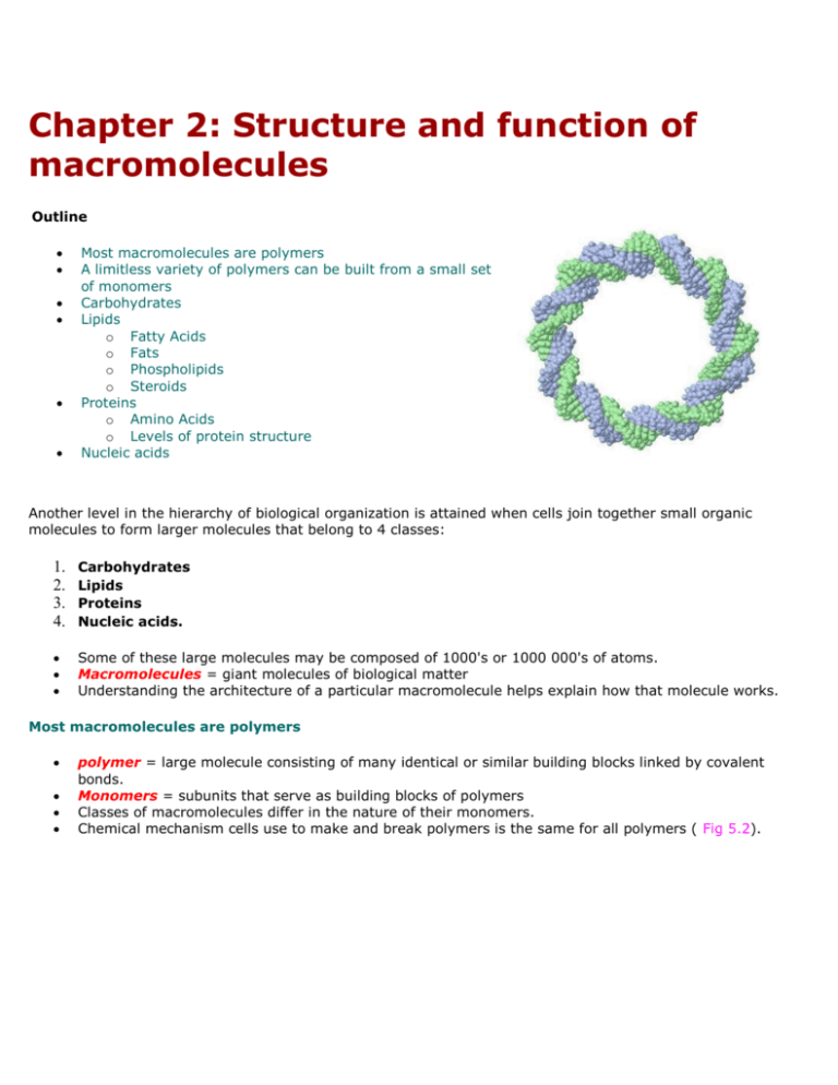
Chapter 2: Structure and function of macromolecules Outline Most macromolecules are polymers A limitless variety of polymers can be built from a small set of monomers Carbohydrates Lipids o Fatty Acids o Fats o Phospholipids o Steroids Proteins o Amino Acids o Levels of protein structure Nucleic acids Another level in the hierarchy of biological organization is attained when cells join together small organic molecules to form larger molecules that belong to 4 classes: 1. 2. 3. 4. Carbohydrates Lipids Proteins Nucleic acids. Some of these large molecules may be composed of 1000's or 1000 000's of atoms. Macromolecules = giant molecules of biological matter Understanding the architecture of a particular macromolecule helps explain how that molecule works. Most macromolecules are polymers polymer = large molecule consisting of many identical or similar building blocks linked by covalent bonds. Monomers = subunits that serve as building blocks of polymers Classes of macromolecules differ in the nature of their monomers. Chemical mechanism cells use to make and break polymers is the same for all polymers ( Fig 5.2). o o Dehydration reactions = covalent bonding of monomers through the loss of a water molecule. Requires energy and enzymes. Hydrolysis = disassembly of polymers into monomers by the reverse process as dehydration, i.e. water is added (e.g. during digestion). A limitless variety of polymers can be built from a small set of monomers All macromolecules are constructed from only 40-50 common monomers. Molecular logic of life is simple yet elegant: Small molecules common to all organisms are ordered into unique macromolecules Carbohydrates Carbohydrates = sugars and their polymers Most abundant biological molecules on earth. Product of photosynthesis. Play several key roles in living organisms: 1. carbohydrates serve as precursors to all other biological molecules. 2. oxidized to yield energy 3. polymers have structural functions - protective coatings, cellulose, chitin etc. 4. derivatives found in other molecules - e.g. ribose in ATP, glycoproteins, glycolipids etc.. - cell-cell interactions and immune system Carbohydrates are described by the number of monomeric units they contain: Monosaccharides o simple sugars o generally have molecular formulas that are multiples of CH2O o names of monosaccharides generally end in "ose". o o o o o can be of varying length (3-7 carbons long). Hexose sugars are most common. some sugars vary only in spatial arrangements of their parts around an asymmetric carbon (e.g. glucose and galactose) in aqueous solutions most sugars from ring structures (Fig 5.4) Monosaccharides serve as major nutrient source for cells. Their carbon skeletons serve as raw material in the formation of other biological molecules. Disaccharides refer to Fig 5.5 o double sugar o depending on location of carbonyl group, sugars can be ketoses (fructose) or aldoses (glucose). Trademarks include 1. hydroxyl group attached to each carbon but one 2. which is bonded to carbonyl group. consists of two monosaccharides joined by a glycosidic linkage e.g. 2 glucoses make maltose 1 glucose + 1 galactose = lactose 1 glucose + 1 fructose = sucrose (table sugar) Polysaccharides o macromolecules consisting of 100's or 1000's of monosaccharides linked together by glycosidic bonds. Storage polysaccharides (refer to fig 5.6) o 1. Starch storage polysaccharide of plants consists entirely of glucose monomers o monomer linked by 1-4 alpha glycosidic linkages amylose = unbranched starch amylopectin = branched starch most animals have enzymes which hydrolyze starch o 2. Glycogen storage polysaccharide in animals (liver and muscles) also made of glucose monomers but is more extensively branched than starch. Both glycogen and starch polymers have a helical shape resulting from their 1-4 alpha glycosidic bonds. Structural polysaccharides (refer to Fig 5.7) o 1. Cellulose major component of plant cell walls most abundant organic compound on earth polymer of glucose, but are linked by 1-4 beta glycosidic linkages (as in cellobiose). This difference gives cellulose a different shape and properties than starch and glycogen. Fig 5.8 many cellulose molecules held together by H-bonding between hydroxyl group of glucose monomers, arranged in units called microfibrils. enzymes that hydrolyze alpha bonds unable to hydrolyze beta bonds very few organisms produce cellulases, enzymes that hydrolyze cellulose. Many fungi use cellulases in their ecological role as decomposers. important source of fiber in human diet. 2. Chitin o structural polysaccharide made from glucose with a nitrogen containing group. major component of arthropod exoskeleton and fungal cell walls. Lipids Consist of hydrophobic molecules with diverse structures and functions. Consist mostly of hydrocarbons (i.e. less oxidized than carbs, therefore have lots of chemical energy) 3 important families of lipids are: 1. Triglycerides 2. Phospholipids 3. Steroids Triglycerides Includes fats and oils. Large molecules (not polymers) constructed from two kinds of molecules ( fig 5.11). o 1. glycerol (3 carbon alcohol with 3 OH groups) o 2. fatty acid (long hydrocarbon with carboxyl group) Hydrocarbon portion of fats make it nonpolar (hydrophobic) 3 fatty acids joined to glycerol by ester linkage ( bond between OH and carboxyl groups). Number of fatty acids determines if its a mono-, di-, or triglyceride. Triglycerides differ in the types of fatty acids attached. Do not have to be the same. Refer to Fig 5.12 Saturated fats = hydrocarbon chains contain maximum number of bonded hydrogen atoms (i.e. no double bonds). Usually solid at room temperature. o Unsaturated fats = one or more double bonds present in carbon skeleton. Double bonds introduce kinks in hydrophobic tail and so these molecules do not pack as well and are usually liquid at RT. We usually call them oils. More predominant in plant sources. Functions of triglycerides o 1. Compact energy storage o 2. Insulation (subcutaneous fat) o 3. Cushions internal organs Phospholipids Structurally related to triglycerides, but have only 2 fatty acids (Fig 5.13). Third OH group of glycerol bonded to a phosphate group (electrically charged). Different phospholipids can differ by what other (usually polar) groups are attached to the phosphate. Have hydrophilic heads and hydrophobic tails (Amphipathic). o Phospholipids in water spontaneously assemble into micelles and phospholipid bilayers (and liposomes). In these structures, the nonpolar, hydrophobic tails are tucked away from contact with water, and the polar, hydrophilic heads of the phospholipids are facing the aqueous environment. Cell membranes are made of phospholipids and are also bilayers ( Fig 5.14). The fact that phosphplipids in water so readily form liposomes with bilayers suggests that if the early earth accumulated phospholipids by chemical evolution, then they would certainly form water filled vesicles, capable of maintaining an internal environment different from the external. These vesicles would eventually become the first cells. Steroids Characterized by carbon skeleton consisting of 4 interconnected rings (Fig 5.15). Cholesterol is a precursor of all steroid hormones (e.g. sex hormones). Also present in cell membranes, where they regulate membrane fluidity. Different steroids differ in functional groups attached. Proteins Most functionally and structurally diverse of the macromolecules. Their functions are fundamental to cellular life. There are many types of proteins varying functions (table 5.1) o 1. Structural proteins (support: e.g. silk, collagen, keratin... etc.) o 2. storage proteins (ovalbumin in eggs, zeins in corn seeds, casein in milk, etc...) o 3. transport proteins (O2 by hemoglobin, ion transporters in cell membrane) o 4. hormonal proteins (coordination of organism's activities: e.g. insulin, glucagon, etc...) o 5. receptor proteins (response of cell to chemical stimuli: e.g. neurotransmitter receptors, hormone receptors, etc...) o 6. contractile proteins (involved in movement, e.g. actin and myosin.) o 7. defense proteins (protection against disease, e.g. antibodies) o 8. Enzymatic proteins (most crucial of functions; selective acceleration of chemical reactions)(Fig 5.16) Each protein has a unique 3-D conformation and therefore often a unique function. Proteins are polymers formed by monomers called amino acids.( Fig 5.17) Amino acids consist of an asymmetric carbon bonded to 4 different covalent partners: Different amino acids differ in their R-group. o R-group determines physical and chemical characteristics of a particular amino acid. R-groups can be nonpolar, polar, or electrically charged (i.e. ionic). Amino acids can be covalently linked through condensation reactions to form polymers. This covalent linkage is called a peptide bond. (Fig 5.18) All polypeptide chains have a polarity: N-terminus and carboxy-terminus. Polypeptides have common backbone of -N-C-C-N-C-C-N-C-C-(N-C-C)nPolypeptides range in size from a few to 1000's of monomers. Unlimited number of polypeptides can be made by varying the number and the order of amino acids in the chain. Protein function depends on its 3 dimensional shape (i.e. its conformation). o the sequence of amino acids along a polypeptide chain determines the conformation of the protein. o correct 3-D folding of polypeptide determines the function of a protein. This is because often protein function depends on recognition and binding of other molecules (binding like a lock and key) (Fig 5.19: lysozyme) Levels of protein structure When cells make a polypeptide, the chain folds spontaneously (i.e. associated with an increase in entropy) to assume the functional conformation of that protein. 4 superimposed levels of structure (Fig 5.21) Primary structure o Order of amino acids along a chain o Primary structure of protein is determined by the sequence of codons in DNA. o Determines other structural levels o Single changes in amino acid sequence may have profound impact on protein function (e.g. Sickle-cell anemia) o Insulin was first protein to be sequenced by Sanger in the late 1940's o Axiom: "each polypeptide has a specific primary sequence" Secondary structure o local coils and folds resulting from H-bonds at regular intervals of polypeptide backbone (i.e. Rgroup not involved in H-bonding). o alpha helix: coil held together by H-bonding between every 4 a.a. pleated sheet: chain folds back in parallel or antiparallel orientation, and H-bonds between parallel regions hold structure together. Tertiary structure o o o Overall 3-D shape of protein Result from irregular contortions from bonding between side chains (R-groups) of various amino acids. Hydrophobic interactions are a strong determinant in protein folding (a.a. with hydrophobic Rgroups congregate at core of protein) o H-bonds, ionic interactions, and disulfide bridges of side chains also involved in stabilizing the tertiary structure. Quaternary structure o Some proteins consist of two or more polypeptide chains. o Quaternary structure is the overall protein structure that results from the aggregation of these polypeptide units e.g. collagen = triple helix (3 subunits) e.g hemoglobin = 2 alpha and 2 beta subunits o In addition to primary structure, protein conformation is also dependent on protein's environment (e.g. changes in temp, pH, of salt concentration can lead to protein denaturation ( unfolding of protein with resultant loss of function) (Fig 5.23) o We still can not fully predict the 3-D conformation by knowledge of primary sequence only. Too complex. o Nucleic acids (genetic material) Nucleic acids encode the genetic information (i.e. primary structure of proteins). Information flow proceeds from DNA to RNA to protein. This is called "central dogma".(Fig 5.25) 2 types of nucleic acids 1. Deoxyribonucleic acid (DNA) - deoxyribose sugar - double stranded (helix) - have thymine rather than uracil 2. Ribonucleic acid (RNA) - ribose sugar - single stranded - uracil instead of thymine Nucleic acid polymers (DNA and RNA) consist of joining together of monomers called nucleotides. The order of nitrogenous bases extending from phosphate-sugar backbone determines amino acid sequence in proteins. DNA consists of 2 chains of nucleotides that spiral an imaginary axis to form a double helix ( Fig 5.28). o Watson and Crick (1953)- determined structure of double helix Strands kept together by H-bonding between complementary bases on each strand (A-T, and C-G). During DNA replication, strands come apart and each acts as a template to synthesize complementary DNA molecules. All nucleotides composed of (Fig 5.26) o 1. phosphate group o 2. pentose sugar o 3 nitrogenous base o Different nucleotides differ in their nitrogenous base. There are two families of nitrogenous bases: Pirimidines: cytosine (C), thymine (T), uracyl (U) Purines: adenine (A), guanine (G) Polymer structure (Fig 5.27b) o o nucleotides are joined by covalent bonds called phosphodiester linkages, between phosphate of one nucleotide and the sugar of next monomer. DNA backbone is a phosphate. Stucture of DNA is ideally suited to its function: encodes information replicates easily grooves allow sequence specific binding of proteins mutates (allows evolution).

