Metastasis powerpoint
advertisement
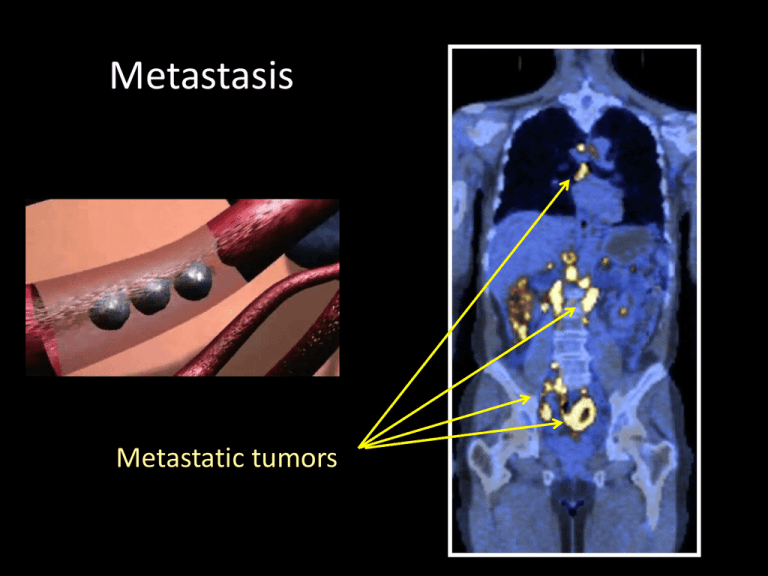
Metastasis Metastatic tumors Figure 20-1 Molecular Biology of the Cell (© Garland Science 2008) Cancer develops through gradual changes in cell morphology and properties benign tumor malignant tumor You may not believe it but by the end of the semester This will make sense! Hanahan and Weinberg, Cell 100:57-70 (2000) Where do they go? Metastatic tropism Figure 14.42 The Biology of Cancer (© Garland Science 2007) Metastatic tropism - Cells find their way to the target tissue via - Metastasis Figure 14.17b The Biology of Cancer (© Garland Science 2007) An organ is composed of several tissues Epithelial cells Connective tissue Muscle tissue Cancer cells need to change their epithelial properties, to lose their adhesion and to penetrate through potent physical barriers basal lamina connective tissue How do they do that? The same way normal cells do it Metastasis Figure 14.17b The Biology of Cancer (© Garland Science 2007) Intravasation Metastasis Figure 14.17b The Biology of Cancer (© Garland Science 2007) The blood: a hostile environment - Cells are normally anchorage-dependent (anoikis) - Shear forces tear cells apart http://www.cancerquest.org/ Figure 14.7b The Biology of Cancer (© Garland Science 2007) Colonization First, micrometasteses Figure 14.10a The Biology of Cancer (© Garland Science 2007) Dormant micrometasteses are viable Figure 14.12 The Biology of Cancer (© Garland Science 2007) Figure 14.50a The Biology of Cancer (© Garland Science 2007) Eventually: macrometastases Intravasation Latency Colonization Steeg Nature Med 06 Angiogenesis Nguyen, Nature Rev. Cancer 2009 Metastatic inefficiency A sequence of inefficient steps Figure 20-44 Molecular Biology of the Cell (© Garland Science 2008) Back to the first steps How do cells become invasive? EMT Epithelial to Mesenchymal Transition sea urchin embryo Figure 14.13a The Biology of Cancer (© Garland Science 2007) EMT Epithelial to Mesenchymal Transition Major changes during EMT - Loss of E-cadherin - Cell shape changes driven by Rho GTPases - MMPs cadherin actin Figure 13.12d The Biology of Cancer (© Garland Science 2007) Adopting changes typical to EMT Figure 14.15b The Biology of Cancer (© Garland Science 2007) Epithelial marker Mesenchymal marker Figure 14.19c The Biology of Cancer (© Garland Science 2007) Rho family proteins promote actin remodeling Svitkina and Borisy JCB 99 MMPs (matrix metalloproteinases) help the cancer cells to invade the ECM Major changes during EMT - Loss of E-cadherin - Cell shape changes driven by Rho GTPases - MMPs Epithelial marker Mesenchymal marker Figure 14.20e The Biology of Cancer (© Garland Science 2007) Figure 14.25 The Biology of Cancer (© Garland Science 2007) Angiogenesis MMP-9 To learn more about the interactions between cancer cells and their microenvironment: 2 review papers posted on your website under “other material” Summary - Invasion-intravasation-circulation-extravasationcolonization - Metastatic cells follow the EMT program - Major changes: cell adhesion, cell shape changes, and secretion of MMPs - Tumor cells rely on stromal cells in their microenvironment - Metastasis is inefficient
