Station 1
advertisement
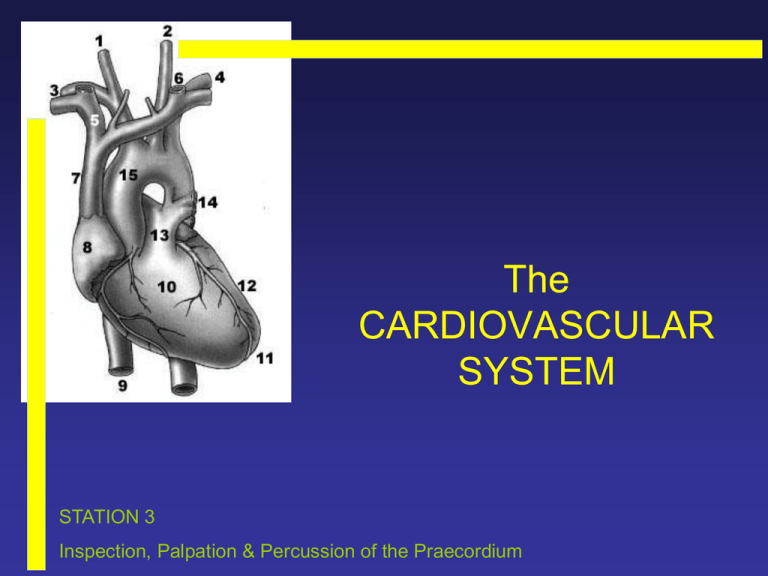
The CARDIOVASCULAR SYSTEM STATION 3 Inspection, Palpation & Percussion of the Praecordium INSTRUCTIONS There are 9 slides in this loop. Work through the slides and practice the examination on the SP. Listen to the recordings on slide 9. Please only move to the next station after the bell has rung. INSPECTION • SHAPE pectus excavatum pectus carinatum barrel-shaped INSPECTION cont. • SCARS Median sternotomy coronary artery bypass surgery valve surgery Thoracotomy mitral valvotomy PALPATION • APEX BEAT 5th intercostal space just medial to MCL; palpate with fingertips well localised area of impulse < size of R5 coin Displaces inferolaterally in ventricular enlargement or as a result of chest deformity, pleural / pulmonary disease Apex beat R5 coin PALPATION • APEX BEAT – the character Pressure loaded: systolic overloaded / hyperdynamic forceful & sustained impulse Volume loaded: diastolic overloaded / hyperkinetic unco-ordinated impulse felt over larger area than usual L ventricular dysfunction Double impulse: two impulses felt with each systole hyprtrophic cardiomyopathy Tapping apex beat: palpable first heart sound mitral stenosis PALPATION cont. • PARARSTERNAL HEAVE place heel of hand just lateral to the left parasternal border right ventricular enlargement & severe left atrial enlargement right ventricle pushed anteriorly & heel of hand lifted off chest wall with each systole • THRILLS Thrills may also be described as palpable murmurs and are as a result of turbulent blood flow. Usually felt in the base and apex of the heart PERCUSSION • A technique you should all be comfortable with by now, can be used to define the cardiac borders. AUSCULTATION • Click on the following numbers below in sequence and listen to the recordings about normal heart sounds. • • • • • • • 1 2 3 4 5 6 7 Introduction Normal 1st & 2nd heart sounds Production & components of 1st sound Normally split 1st sound Production & components of 2nd sound Fourth heart sound Third heart sound

