Best Practice in Cryosurgery Presentation
advertisement
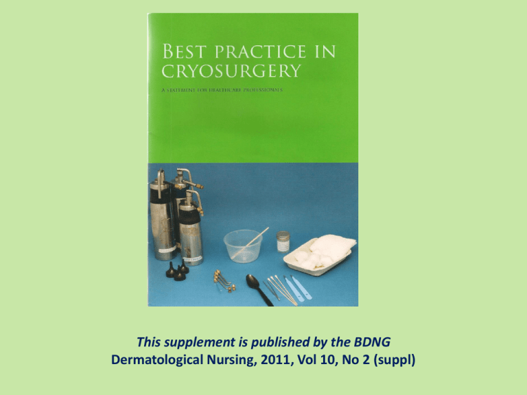
This supplement is published by the BDNG Dermatological Nursing, 2011, Vol 10, No 2 (suppl) Cryotherapy – Introduction Cryotherapy: the destruction of skin lesions using a cold substance most commonly liquid nitrogen LN2 (-196°C; -321°F) destruction is selective, affecting tissue only induction of an effective immune recognition of viral or tumor cells Cryotherapy - Indications Treatment of: Benign lesions Premalignant lesions Malignant lesions Table 1. Some of the common conditions responsive to cryosurgery. Benign lesions 8 Viral Warts 8 Skin tags 8 Seborrhoeic keratoses 8 Sebaceous hyperplasia 8 Molluscum contagiosum 8 Milia Pre-malignant lesions 8 Actinic/solar keratoses 8 Bowens disease (Intra-epithelial carcinoma) 8 Actinic cheilitis Malignant Lesions 8 Superficial basal cell carcinomas Cryotherapy - Contraindications There are no absolute contraindications Caution is needed when treating the following conditions: Table 2. Contraindications (Jackson et al, 2006) 8 8 8 8 8 8 8 8 8 8 8 Agammaglobulinaemia Cold intolerance Cold Urticaria Concurrent treatment with renal dialysis Collagen and autoimmune disease The immunosuppressed Cryoglobulinaemia Multiple myeloma Platelet deficiency disease Pyoderma gangrenosum Raynaud’s disease Cryotherapy - Equipment Table 3. Equipment required for cryosurgery 8 8 8 8 8 8 8 8 8 8 8 8 8 8 8 8 Cryospray with a selection of nozzles Flasks Guards Auroscope earpieces/open cones Cotton wool balls/orange sticks Plastic Teaspoon Metal forceps Disposable scalpels (size 15) Gallipot/Styrofoam cup Magnifying glass/Dermatoscope Examination light Non-sterile gloves Aprons Sharps disposal bin Suitable dressings Gauze The equipment required depends on the method and technique used Methods: Open spray - 40 ̊C Cotton bud - 20 ̊C Metal forceps - 15 ̊C Cryotherapy - Methods Spray: Spot freeze most commonly used for lesion up to 2cm diameter Table 4. Rim of normal skin (Andrews MD, 2004). Benign lesions 1-2mm Premalignant lesions 2-3mm Malignant lesions* 5mm Superficial BCC Actinic keratosis and Bowens Do not use cryosurgery to treat lentigo maligna *Reminder: melanocytic lesions are not suitable for cryosurgery If lesion is over 2cm diameter use paint brush or rotary/spiral technique Include rim of normal tissue Cryotherapy - Techniques A freeze / thaw cycle is used Following the freezing the lesion must thaw fully as this is part of the cell destruction Spray – 1cm distance from lesion Freeze time starts from when the area is white Once ice has developed continue for an appropriate time of 5-30 seconds intermittently Cryotherapy - Procedure Table 5. Procedures for administration. The following procedures must be used in conjunction with local policies and procedures. Action Rationale General patient assessment and skin surveillance including lesion assessment To assess suitability of both patient (age and mental capacity) and lesion for treatment Provide verbal and written explanation of the procedure to the patient/guardian/carer and gain informed consent To ensure the patient fully understands what the treatment involves and to elicit concordance and reduce anxiety Assess lesion(s) for treatment and document; this could involve: measurements, photographs, body maps, diagrams Essential for monitoring efficacy of treatment and record keeping Hyperkeratotic lesions should be pared down prior to treatment See Appendix 2 Keratin can act as an insulator and can make the treatment less effective Select and prepare appropriate equipment To ensure that treatment is delivered in a safe, effective and efficient manner Cryotherapy - Procedure Table 5. cont. Procedures for administration. The following procedures must be used in conjunction with local policies and procedures. Action Rationale Spray technique This is to ensure maximum effect and best possible clinical outcome from procedure Select appropriate nozzle according to the size of the lesion Hold tip 1cm from the skin over the centre of the lesion Spray gently until lesion and 1mm-5mm rim of healthy tissue becomes white (frozen) Some patients may have low tolerance to cryosurgery and may experience extreme reactions to treatment ranging from discomfort to pain. Therefore in some cases it may be advisable to administer a test dose on the lesion Maintain freeze with intermittent spraying for 5-30 seconds as appropriate Allow to thaw Repeat with second freeze as above if necessary If indicated apply topical steroid as prescribed Application of topical steroids may reduce post inflammatory reaction particularly in facial areas Cryotherapy - Procedure Table 5. cont. Procedures for administration. The following procedures must be used in conjunction with local policies and procedures. Action Rationale Cotton bud technique This is to ensure maximum effect and best possible clinical outcome from procedure Prepare applicator using orange stick and cotton wool The area of the tip of the cotton wool should be slightly smaller than the area to be treated To ensure the applicator is tailored to the individual lesion therefore minimising trauma to healthy surrounding skin Decant a small amount of liquid nitrogen into a non-metallic vessel such as a gallipot or a Styrofoam cup. The flask should never be used as the reservoir due to risk of crosscontamination A fresh cotton bud and vessel must be used for each patient, otherwise contamination will occur. Viruses such as human papilloma virus, herpes virus and hepatitis strains can remain viable at temperatures as low as -196⁰C The cotton bud is dipped into the liquid nitrogen for a minimum of 10 seconds Immediately apply firmly and vertically to lesion Continue until whole lesion and appropriate margin is frozen Repeat as above if required to maintain an appropriate ice field The liquid nitrogen should be allowed to evaporate prior to disposal of vessel To prevent thermal injury to practitioner Only suitable for small, superficial lesions Useful when treating young children Cryotherapy - Procedure Table 5. Procedures for administration. The following procedures must be used in conjunction with local policies and procedures. Action Rationale This is to ensure maximum effect and best possible Forceps method clinical outcome from procedure Wrap gauze around the handle of the metal forceps To prevent thermal injury to practitioner Decant a small amount of liquid nitrogen into a nonmetallic vessel such as a gallipot or a Styrofoam cup A fresh pair of forceps and vessel must be used for each patient, otherwise contamination will occur. Viruses such as human papilloma virus, herpes virus and hepatitis strains can remain viable at temperatures as low as 196⁰C Dip the forceps into a vessel filled with liquid nitrogen and leave until it becomes frosted (this will take approximately one minute) Grasp the lesion, including the base, and pinch until ice ball is formed and keep in place for 5-10 seconds Repeat as above if required to maintain an appropriate ice field For resistant lesions this technique can be used in conjunction with the spray method to the base If required, cover treated lesions with sterile, dry dressing To reduce the risk of infection if skin is broken Patient may like the area protected following treatment Give patient written after-care advice and recommended adjunct therapies To ensure that patients can self-manage minor expected adverse effects and how/where to seek help Cryotherapy - Documentation Table 5. Documentation Action Rationale Document: 8 Effects of previous treatments (if any) 8 Lesion treated 8 Technique used 8 Nozzle size if appropriate 8 Number of freeze-thaw cycle 8 Length of freeze time 8 Patient tolerability 8 Adverse events 8 After-care instructions/adjunct therapies A checklist may be useful. See example in Appendix 3 Records of treatment should be completed with the patient present if possible to ensure agreement. They should be clear and accurate to allow them to be interpreted by others. To standardise practice between practitioners Cryotherapy - Indications Table 6. Indications for cryosurgery Benign lesions Pre-malignant lesions Malignant lesions (under the care of specialist dermatology services) 8Viral warts 8Skin tags 8Seborrhoeic keratoses 8Sebaceous hyperplasia 8Molluscum Contagiosum 8Milia 8 Actinic/solar keratoses 8 Bowen’s disease (Intraepithelial carcinoma) 8 Actinic cheilitis 8 Superficial basal cell carcinomas Cryotherapy - Viral Warts Table 7. Common forms of viral warts Clinical type Appearance Common Firm, rough papules and nodules on any skin surface. May be single or grouped 2-4 mm in diameter, slightly elevated but most commonly flat-topped papules with minimal scaling Have features of common and plane warts Plane (flat) Intermediate Myrmecia (verruca) Deep burrowing warts Plantar May start as ‘sago grain-like’ papules, which develop a more typical keratotic surface with a collar of thickened keratin Occur when palmer and plantar warts coalesce into larger plaques Mosaic Cryotherapy – Viral Warts Viral warts: Infection of the epidermis Human papilloma virus (HPV) Treatment: Either with spray or cotton bud A bud should be used on the face Two freeze thaw cycles are recommended Cryotherapy – Skin Tags Skin Tags: Common, soft, harmless lesion of collagen and blood vessels Treatment: Forceps method or spray method For spray method hold tag away from skin and freeze through the base of tag Cryotherapy – Seborrhoeic Warts Seborrhoeic Warts: Common benign lesions often starting in adulthood Warty, waxy, stuck on appearance Cryotherapy – Molluscum contagiosum Molluscum contagiosum : Small (1-5mm) lesions caused by pox virus infection of the skin Common in : Atopic eczema Immunocompromised patients Children Treatment: Spray method Cryotherapy – Sebaceus Hyperplasisa Sebaceous Hyperplasisa : Enlarged sebaceous glands Common in : Middle aged or elderly patients Immunocompromised patients Torré-Muir syndrome sebaceous gland tumor Treatment: Spray method Cryotherapy – Millia Millia: Tiny, superficial, keratin filled epidermal cysts Common in : Infants and adults Congenital or acquired Can result from physical trauma Sebaceous or sweat duct plugging Treatment: Spray method Cryotherapy – Actinic/Solar Keratoses Actinic kerartosis: Hyperkeratotic lesion, chronic sun damage Pink, scaly, warty or crusted lesion Common in : Adult skin Light skinned individuals Treatment: Spray method Cryotherapy – Bowen’s disease Bowen’s disease: Persistent, non-elevated, red, scaly or crusted plaque Has small potential for invasive malignancy to SSC Can grow several cm in diameter Common in : Elderly patients Lower legs Treatment: Spray method Actinic Cheilitis Actinic Cheilitis BCC Superficial BBC Cryotherapy – Side effects Pain Oedema/Blister Ulceration Nerve/tendon damage Pigment change Scarring Infection Urticaria Cryotherapy - Safety Precautions Storage Liquid nitrogen should always be stored in a well ventilated room. aware of potential hazards, Especially asphyxiation, and know what to do in the event of an accident or emergency (BOC, 2004). Personal protective equipment When decanting liquid nitrogen non absorbent insulated gloves and a full face visor should be worn. Open toed shoes should not be worn It should only be transported where the load space is separated from the driver and passenger compartment. Transportation If liquid nitrogen is to be transported in a vehicle, the driver must be Liquid nitrogen containers should be • transported in a secure upright position • in a well-ventilated area (BOC, 2004). Cryotherapy - Safety COSHH regulations apply Hazards include: Asphyxiation in poorly ventilated areas Chronic burns Cryogenic burns / frost bite Hyperthermia Wear protective equipment when handling Emergency action: Inhalation: Remove individual from area Do not place yourself at risk Breathing apparatus may be used Keep individual warm Skin/Eye contact: Immerse affected area in tepid 42-45°C for at least 15min and cover with dry, sterile dressing Cryotherapy – also covers… Medicolegal aspects Appendix 1: Assess competency according to WASP framework Appendix 2: Methods for removal of keratin Appendix 3: Check list for cryotherapy Dermatological Nursing, 2011, Vol 10, No 2 (suppl) BDNG Tel: 020 7681 6131 www.bdng.org.uk
