Folie 1 - Angiopro
advertisement
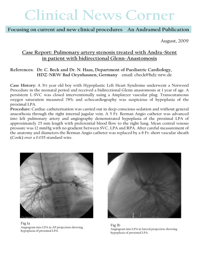
Clinical News Corner Focusing on current and new clinical procedures An Andramed Publication August, 2009 Case Report: Pulmonary artery stenosis treated with Andra-Stent in patient with bidirectional Glenn-Anastomosis References: Dr. C. Beck and Dr. N. Haas, Department of Paediatric Cardiology, HDZ-NRW Bad Oeynhausen, Germany email: cbeck@hdz-nrw.de Case History: A 3½ year old boy with Hypoplastic Left Heart Syndrome underwent a Norwood Procedure in the neonatal period and received a bidirectional Glenn anastomosis at 1 year of age. A persistent L-SVC was closed interventionally using a Amplatzer vascular plug. Transcutaneous oxygen saturation measured 78% and echocardiography was suspicious of hypoplasia of the proximal LPA. Procedure: Cardiac catheterisation was carried out in deep conscious sedation and without general anaesthesia through the right internal jugular vein. A 5 Fr. Berman Angio catheter was advanced into left pulmonary artery and angiography demonstrated hypoplasia of the proximal LPA of approximately 25 mm length with preferential blood flow to the right lung. Mean central venous pressure was 12 mmHg with no gradient between SVC, LPA and RPA. After careful measurement of the anatomy and diameters the Berman Angio catheter was replaced by a 8 Fr. short vascular sheath (Cook) over a 0.035 standard wire. Fig 1a Angiogram into LPA in AP projection showing hypoplasia of proximal LPA. Fig 1b Angiogram into LPA in lateral projection showing hypoplasia of proximal LPA. A 21 mm XL AndraStent (Andramed Germany) was hand-crimped on a 7 mm x 20 mm Opta Pro Ballon (Cordis) and hand injections confirmed optimal stent position in the proximal LPA. The balloon was inflated with 10atm using an inflator (Fig 3). To archieve equal vessel diameter in both right and left proximal pulmonary artery we re-dilated the stent with a 9 mm x 20 mm Opta Pro Ballon (Cordis) (Fig. 4). The balloon was inflated with 10 atm using an inflator. The post implantation angiogram demonstrated an optimal stent position, no extravasation and improved flow to the left lung (Fig. 5, 6). The stent shorting was 4% with a homogeneous diameter. Overall procedure time was 31 minutes with a fluoroscopy time of 5 minutes. The patient was discharged home 48 hours post procedure. Fig 3 Fig 4 Angiogram after stent implantation in AP projection. Picture of stent re-dilation in AP projection. Fig 5 Fig 6 Angiogram after stent re-dilation showing increased proximal LPA diameter in AP projection . Picture of stent after re- dilatation in AP projection. Result: Stent placement through a short vascular sheath was easily achievable in this patient with Glenn anastomosis. Re-Dilation of the Andra Stent stent was easily done to achieve an optimal anatomic and excellent angiographic result. Together with the added benefits of the chromium-cobalt technology as well as the semi-open cell design, placing the stent in a angulated part of pulmonary vessels seems easily achievable in patient with univentricular physiology.
