Respiratory System Ppt
advertisement
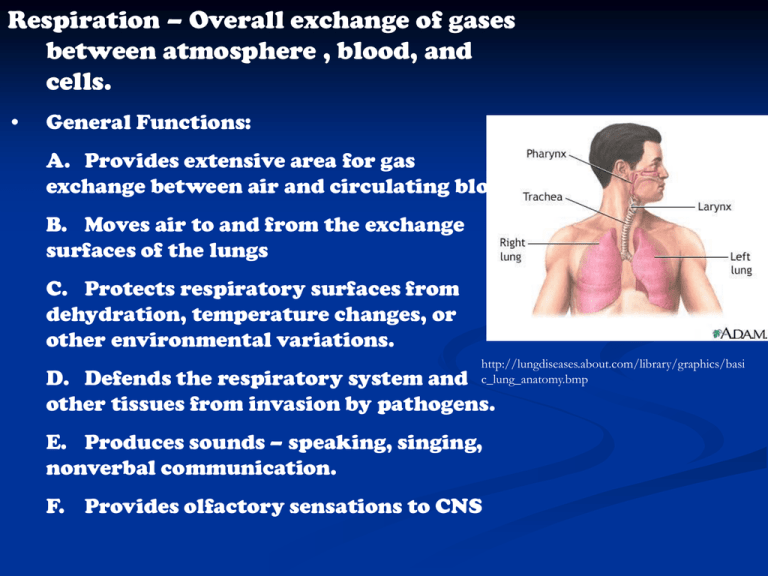
Respiration – Overall exchange of gases between atmosphere , blood, and cells. • General Functions: A. Provides extensive area for gas exchange between air and circulating blood B. Moves air to and from the exchange surfaces of the lungs C. Protects respiratory surfaces from dehydration, temperature changes, or other environmental variations. http://lungdiseases.about.com/library/graphics/basi c_lung_anatomy.bmp D. Defends the respiratory system and other tissues from invasion by pathogens. E. Produces sounds – speaking, singing, nonverbal communication. F. Provides olfactory sensations to CNS II. Processes involved in respiration: A. Pulmonary Ventilation – (Breathing) The physical movement of air into and out of the lungs (more specifically the alveoli) B. External Respiration – Exchange O2 and CO2 between interstitial fluids and the external environment. C. Internal Respiration – Absorption of O2 and release of CO2 by the cells (cellular respiration in the mitochondria) http://www.apparelyzed.com/_images/content/respri tory/Respiratory-System.jpg http://biology.clc.uc.edu/graphics/bio105/respiratory.jpg III. Organs of respiration: A. Respiratory Tract – airways that carry air to and from exchange surfaces 1. Conducting portion – lined with respiratory mucosa, respiratory defense system – sticky mucus, filtration, alveolar macrophages 2. Respiratory portion – site of gas exchange B. Upper Respiratory Tract – nose, nasal cavity, paranasal sinuses, pharynx – filter, warm, humidify incoming air and dehumidify and cool outgoing air 1. Nose a. External nares - nostrils b. Nasal Cavity – highly vascularized, divided by nasal septum, as exhale heat is absorbed by blood vessels and moisture condenses which prevents drying http://www.askdrs hah.com/images/na sal_polyp.jpg c. paranasal sinuses – mucous secretions and tears keep surfaces clean and moist, olfactory receptors, nasal conchae cause air to eddy and swirl which traps foreign objects, warms and humidifies air. 2. Pharynx – Chamber shared by digestive and respiratory systems, divided into: a. nasopharynx – superior, pharyngeal tonsil, auditory tubes open into b. oropharynx – between soft palate and base of tongue, palatine tonsils c. laryngopharynx – between hyoid and entrance to the larynx and esophagus, resists abrasion, chemical attack, pathogens (stratified squamous epithelium) C. Lower Respiratory Tract – larynx, trachea, bronchi, bronchioles, alveoli 1. Larynx – voice box, located at C4 or C5 to C6 a. Glottis – narrow opening to larynx b. Made up of nine cartilages 3 large unpaired: Thyroid (shield-shaped) – largest, hyaline, U-shaped, incomplete posteriorly (Adam’s Apple) Cricoid (ring-shaped) - hyaline Epiglottis – elastic , forms lid over glottis during swallowing 3 paired: Arytenoid, Corniculated (sound), Cunieform http://academic.kellog g.cc.mi.us/herbrandso nc/bio201_McKinley/f 25-4_larynx_c.jpg http://academic.kellogg.cc.mi.us/herbrandsonc/bio201_M cKinley/f25-4_larynx_c.jpg http://www.gbmc.org/bin/r/n/Larynxc utsm.jpg c. VOCAL FOLDS (CORDS) – Highly elastic, protected by VESTIBULAR FOLDS (false vocal cords), when air passes over, vibrates to produce sound waves - PHONATION PITCH – Determined by diameter, length (larynx size) and tension (voluntary) ARTICULATION – modifying sounds with pharynx, oral cavity, nasal cavity, sinuses, tongue, lips, cheeks COUGHING REFLEX – Food touches vestibular or vocal cords http://notsosynonymous.tripod.com/images/cords.jpg 2. Trachea – windpipe a. tough, flexible tube, 2.5 cm in diameter and 11 cm long b. located anterior to C6 – T5 c. tracheal cartilages – bound to neighboring cartilage which stiffens to prevent collapse or overexpansion, Cshaped permits posterior trachea to distort for swallowing d. Intubation e. Tracheostomy http://www.sdhct.nhs.uk/patientCare/pil/images /tracheostomy2.jpg http://www.aic.cuhk.edu.hk/web8/Hi%2 0res/Intubation.jpg f. The trachea branches into the left and right PRIMARY BRONCHI (sing. Bronchus) http://www.nlm.nih.gov/medlineplus/ency/images/e ncy/fullsize/1103.jpg g. The primary bronchi enter the lungs 3. Lungs a. Divided into lobes which are separated by fissures b. Located in pleural cavities separated by the mediastinum, hilus allows access for pulmonary vessels and nerves Parietal pleura – lines inner surface of thoracic wall Visceral pleura – covers outer lungs Both pleura secrete pleural fluid – moist, slippery, provides lubrication Pleurisy – lack of pleural fluid http://www.yourlungheal th.org/lung_disease/copd/ healthy/graphics/pleura.j pg http://upload.wikimedia.org/wikipedia/c ommons/thumb/9/9e/Lungs_diagram_sim ple.svg/483pxLungs_diagram_simple.svg.png C. Comparison of left and right lungs Right lung Left lung 3 lobes – superior, middle, inferior 2 lobes – superior, inferior Broader and shorter because diaphragm rises to accommodate liver Longer Indented at cardiac notch http://academic.kellogg.cc.mi.us/herbrandsonc/bio201_McKinley/f2512a_gross_anatomy_o_c.jpg 4. Pathway a. Bronchial Tree Trachea Primary Right Bronchus Primary Left Bronchus (Enter the lungs and divide into) Secondary Bronchi – enter each lobe (3 right, 2 left) Which divide into: Tertiary Bronchi Terminal Bronchioles (6500) Respiratory Bronchioles http://training.seer.cancer.gov/module_anatomy/ images/illu_bronchi_lungs.jpg Alveolar sacs http://academic.kellogg.cc.mi.us/herbrandsonc/bio201_McKinley/f 25-8_bronchial_tree_c.jpg b. Changes in diameter 1. Bronchodilation – Increase in diameter, sympathetic response 2. Bronchoconstriction – Decrease in diameter, parasympathetic, due to allergies, anaphylaxis http://www.asthmameds.ca/images/lungsbig.jpg 5. Microanatomy a. Alveolar sacs are connected to multiple alveoli, surrounded by capillaries b. Alveolar/capillary membrane is composed of three parts: *Squamous epithelial cells lining the alveoli *Endothelial cells lining the capillaries *Basement membrane c. Diffusion occurs quickly across due to: *small total distance separating air from blood *O2 and CO2 are lipid soluble d. SURFACTANT – an oily secretion that decreases surface tensionso alveoli don’t collapse. If not enough is produced, takes a huge amount of effort to inflate the lungs = Respiratory Distress Syndrome http://content.revolutionhealth.com/contenti mages/media-medical-hw-h5550999_001.jpg http://www.mydr.com.au/content/images/categories/lungs/lungs_anatomy.gif


