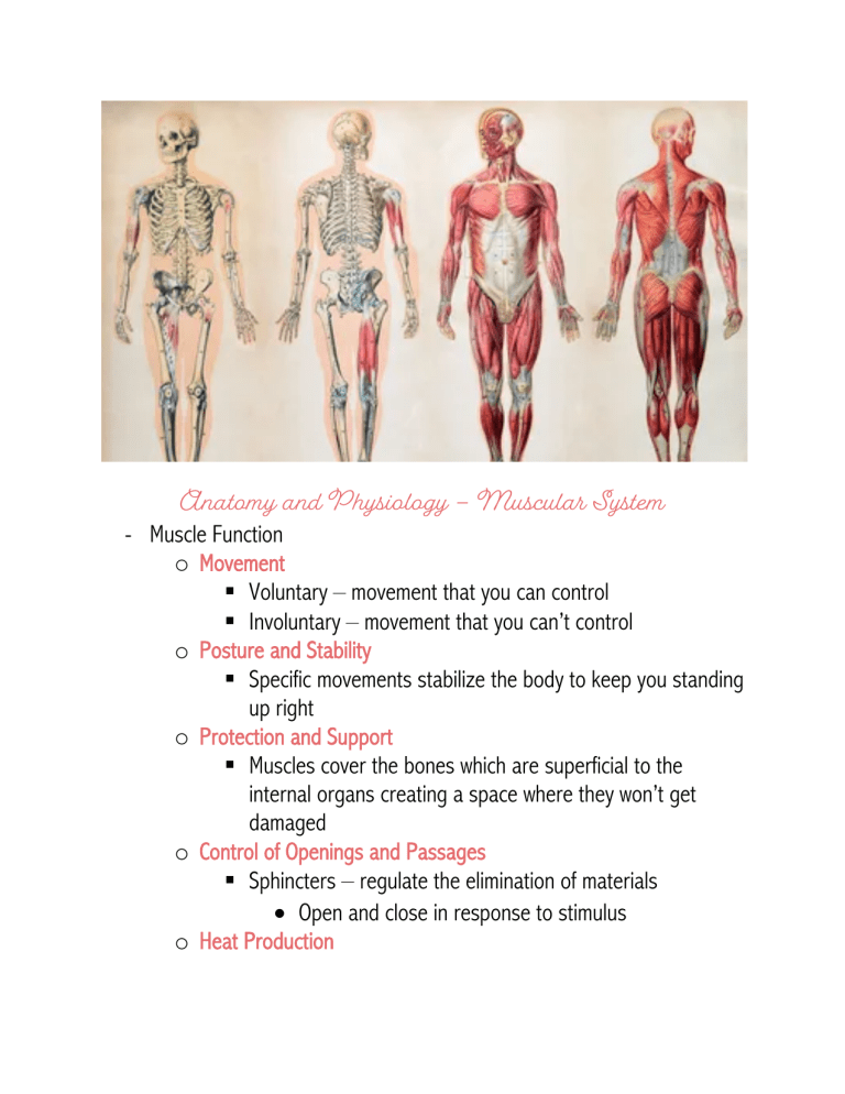
Anatomy and Physiology – Muscular System - Muscle Function o Movement § Voluntary – movement that you can control § Involuntary – movement that you can’t control o Posture and Stability § Specific movements stabilize the body to keep you standing up right o Protection and Support § Muscles cover the bones which are superficial to the internal organs creating a space where they won’t get damaged o Control of Openings and Passages § Sphincters – regulate the elimination of materials • Open and close in response to stimulus o Heat Production § Energy is required for muscles to contract which produces heat. o Control of Glucose § Muscles use energy when contracting which calls for loads of glucose -------------------------------------------------------------------------------------------- Muscular Tissue o Skeletal Muscle – main focus! § Appearance: long parallel cells that are straited (parallel) § Nuclei: multinucleated; usually near the plasma membrane § Control: voluntary o Cardiac Muscle § Appearance: branched network of cells in straited (parallel) layers § Nuclei: uninucleate or binucleate; centrally located § Control: involuntary o Smooth Muscle § Appearance: long cells tapered at the end; no striations § Nuclei: uninucleate; centrally located § Control: involuntary - Skeletal Muscle Composition o Skeletal muscles are organs composed of multiple tissue types (4) § Connective Tissue, Blood Vessels, Nervous Tissue, and Muscular Tissue o Skeletal muscles are built from a simple to complex component § Myofilaments – most basic • Thick Filaments • Thin Filaments § Sarcomere § Myofibril § Muscle Fiber § Fascicle § Muscle – most complex (organ) -------------------------------------------------------------------------------------------- Myofilaments o Myofilaments come together to form myofibrils § Myofilaments are composed of thick and think filaments • Thick – made of myosin • Thin – made of actin, tropomyosin, and troponin § Filament interactions are essential for muscle contraction! -------------------------------------------------------------------------------------------- Myofilaments – Thick Filaments o Myosin Structure § Composed of: • Filamentous Tails • Head o Binding sites for ATP and Actin to interact with myosin § Flexed myosin head = ATP molecule bound § Extended myosin head = hydrolyzed ATP molecules which created ADP and Phosphate • This confirmation allows actin to interact with myosin! § Myosin must be flexible (in two regions) so that it is able to grab and pull molecules § Myosin interacts with other myosin molecules in two ways • Side to side • End to end § Myosin “bundles” = 300 proteins interacting • In the bundle the heads of the proteins are oriented in opposite directions so that during contraction there is cohesive movement in shortening the fibers -------------------------------------------------------------------------------------------- Myofilaments – Thin Filaments o Actin Structure § Actin • Globular Actin (g-actin) – small circle molecule that contains a myosin binding site • Filamentous Actin (f-actin) – assembly of multiple gactin o “beads on a string” § Tropomyosin – wraps around f-actin blocks myosin binding site § Troponin – multi subunit that keep tropomyosin in place on the actin filaments • Binding site for tropomyosin • Binding site for actin • Binding site for calcium o Contraction § In a resting muscle, the actin and myosin interaction can’t happen because the tropomyosin is blocking the binding site § When a muscle contracts the tropomyosin slides off of the myosin binding site and allows the thick and thin filaments to complete contraction -------------------------------------------------------------------------------------------- Sarcomere o Myofilaments are put together to form sarcomeres – the functional unit of muscles § Thick and thin filaments overlap o Z lines/discs are proteins that separate one sarcomere from the next. § Thin filaments are connected directly to the z-lines § Thick filaments are connected to the z-lines by a connectin o M lines are at the midline of the sarcomere and keep the thick filament aligned during contraction o A bands are dark areas which contain the entire thick filament. § Anisotropic – light has a hard time passing through § Slight overlap of thick and thin filaments o H zones are in the A bands and only contain the thick filaments. o I bands contain think filaments and connectin § The I bands spread across multiple sarcomeres and are bisected by the z discs o Every myofibril is enclosed by a sleeve or net of sarcoplasmic reticulum. § Calcium is enclosed here because it always needs to be around for contraction (it unlocked the myosin binding site in actin). - Multiple Sarcomeres o Multiple sarcomeres must contract to make enough tension in a muscle. § In contraction, the thin filaments slide over the thick filaments which shortens the sarcomeres • The A band stays the same length because only the sarcomere length changes not the thick filament length • The H and I band disappear because the thin filaments are being pulled inward to produce contraction -------------------------------------------------------------------------------------------- Skeletal Muscle Fibers o Skeletal muscles are for contraction and force generation – there are many components that make it up § Sarcoplasmic reticulum is the “smooth er” of the muscle fibers • It looks like a net • At the end of each section there are swellings called terminal cisternae (which stores calcium) § Sarcolemma (plasma membrane) • Separation of the extracellular and intracellular environments o Holes in the membrane are from the transverse -tubules) extending into the fiber § Triad – 3 components • T – tubule in the middle of two terminal cisternae • Critical for electrical excitation and mechanical action of muscle fiber o Electrical excitation is a signal that causes movement § Other features of a skeletal muscle cell • Multinucleate • Abundant mitochondria - Myofibril Composition o Skeletal muscle fibers are packed with specialized organelles called myofibril § 80% of the skeletal muscle is composed of myofibrils § Hundreds to thousands per fiber § Extend the entire length of fiber § Each myofibril is enclosed by a sleeve or net of sarcoplasmic reticulum -------------------------------------------------------------------------------------------- Muscle – the organ o Muscles are composed of multiple cells types that are organized into a functional unit by connective tissue § Connective tissue § Blood vessels § Nervous Tissue § Muscular Tissue o Fascicles – groups of skeletal muscle fibers together o Endomysium – connective tissue surrounding single fascicles o Perimysium – dense connective tissue that surrounds fascicles and contain blood vessels and nerves o Epimysium – connective tissue that holds an entire group of fascicles -------------------------------------------------------------------------------------------- Muscle Fiber Characteristics o The function of the muscle (excitation) as an organ is determined (in part) by the properties of the muscle fibers: there are 4 characteristics. § Excitability – muscle cells respond to a stimulus and pass the message down the membrane • Message = A motor neuron • Stimulus is a neurotransmitter (AcH) • Message passed by movement of ions across membrane § Contractibility – shortening of muscles to produce body movement § Extensibility – muscle cells are able to stretch § Elasticity – muscle cells are able to return to their original position after stretching -------------------------------------------------------------------------------------------- Membrane Potential o All cells have a resting membrane potential – a charge difference across the membrane § Membrane potential • The charge difference is due to the unequal distribution of ions. • The resting membrane potential (RMP) is established and maintained by the sodium potassium pump • More Na+ outside the cell and more K+ inside the cell o 2 K+ in and 3 Na+ out • More positive ions leaving the cell causes the intracellular fluid to be more negative o The RMP of a muscle cell is -90mv o The RMP of a neuron is -70 mv -------------------------------------------------------------------------------------------- Voltage and Chemically Gate Channels o Chemically Gated (Regulated) Channels § Open in response to chemical (stimuli) that it associates with • Stimulus = neurotransmitters (ACh) o ACh doesn’t move through ions do (Na+ and K+) § First to open during muscle excitation! Ions move down their concentration gradients • Sodium goes into the cell instead of out • Potassium goes out of the cell instead of in • This makes the charge of the cell go down which impacts the voltage gated channels o Voltage Gated (Regulated) Channels § These channels open and close in response to different voltages which depend upon the changes created by the chemically gated channels § There are multiple voltage gated channels in the membrane § Openings of the channel allow ions to move down the concentration gradient which change the distribution of ions and voltage • Sodium goes into the cell instead of out • Potassium goes out of the cell instead of in o In both cases, open channels permit ions to flow and reach their equilibriums -------------------------------------------------------------------------------------------- Ion Charge Distribution Terminology o The RMP of a muscle cell is -90mV § Depolarization – positive ions (Na+) rush into the cell causing the millivolts of the RMP to become less negative (more positive) • If enough sodium rushes into the cell, then the voltage gated channels will open • Once the voltage reaches +30mV, the Na+ channels close, and repolarization begins § Repolarization – positive ions (K+) rush out of the cell and the cell becomes more negative • To begin repolarization, the K+ channels open and potassium will leave the cell which brings the RMP down from +30 mV to -90mV. -------------------------------------------------------------------------------------------- - Muscular Electrical Signals o Muscles are stimulated to contract by electrical signals that come from neurons § Motor Unit • Somatic motor neurons make electrical signals in the brain or spinal cord o Supply muscles with nerves over a wide area • Somatic motor fibers extend into the muscle and carry these impulses from motor neurons o The motor neurons branch to supply multiple muscle fibers, but each fiber has ONE neuron • The number of motor units will vary o One may control less than 5 muscle fibers which allow precision and less forceful movement o One may control thousands of muscle fibers which allow less precise and higher forceful movements -------------------------------------------------------------------------------------------- - Neuromuscular Junction ” o The area where a motor neuron meets the skeletal muscle cell is called the neuromuscular junction. § Each muscle is stimulated by only ONE neuromuscular junction which is typically in the middle so that the signal can travel down each side (right and left) of the cell • The neuromuscular junction doesn’t physically touch the muscle cell which creates the space between them called the synaptic cleft. o Interaction between the two – § When the neuron stimulates (stimulus = nerve impulse) the muscle cell the synaptic knob takes in calcium which allows it to release ACh which is then released to the synaptic cleft. § When the ACh is released, it ends up binding to the ACh receptors along the sarcolemma. -------------------------------------------------------------------------------------------- Excitation o Excitation (generation of an action potential) of the skeletal muscle begins at the neuromuscular junction when Ach binds to the Ach receptors § Phase 1: • The action potential moves down the neuron which causes calcium to flow into the synaptic knob • When the calcium enters the synaptic knob the vesicles inside the synaptic knob release ACh which enters the synaptic cleft (space between the neuron and the muscle fiber) • When the ACh gets into the synaptic cleft it then binds to the ACh receptors (chemically gated channels) and opens them allowing sodium to flow into the cell (down its concentration gradient) from the extracellular space causing the membrane to reach its threshold (-65mV) § Phase 2: • The action potential that was started by the neuromuscular junction then spreads across the membrane of the muscle fiber to get myofilaments prepared to contract • The sodium continues to flow into the cell until the resting membrane potential is +30mV o This is known as local depolarization • When the membrane potential reaches +30mV the voltage gated channels will open and potassium flows into the cell from the extracellular space which brings the membrane potential back to -90mV (repolarization) -------------------------------------------------------------------------------------------- Excitation – Contraction Coupling o During excitation contraction coupling, action potentials move down the membrane in both directions from the NMJ § Depolarization follows quickly after repolarization o The triad is “shocked” by the action potential (comes from the NMJ) which causes the t-tubules (the middle of the triad) to release calcium (contained in the sarcoplasmic reticulum) from its calcium channels into the cytosol -------------------------------------------------------------------------------------------- Contraction o When calcium gets into the cytosol it prepares the myofilaments for contraction o This stage is dependent upon cross bridge cycling and the proper conformation (set up) of proteins in the sarcomere § Flexed head = ATP molecules bound § Extended head = Hydrolyzed ATP which becomes ADP and Phosphate • This is the energized confirmation which means that it is read to interact with actin! • Once the myosin head is flexed and the myosin and actin interact • This interaction makes the myosin lose the ADP and Phosphate which is called the power stroke. • Once this happens myosin has to detach from actin so that this process is able to repeat -------------------------------------------------------------------------------------------- - Relaxation o The final phase of muscle contraction is relaxation § This processes stops the stimulus, drops calcium levels, and the fibers no longer contract • Nerves fibers stop firing • When the fibers stop firing there is no more ACh releasing, and the remaining is broken down by AChE • Lack of jolting allows the sarcoplasmic reticulum to reabsorb calcium from the cytoplasm (Ca+ ATPase) • Calcium releases from the troponin and the tropomyosin slides back in to place and locks the myosin binding sites -------------------------------------------------------------------------------------------- Clinical Correlate – Neuromuscular Toxins o Neuromuscular toxins interfere with synaptic function and can stop the muscles from contracting § Pesticides contain chemicals that stop AchE from being able to put an end to muscle excitation • Produces spastic paralysis – muscle contraction is unable to be stopped • This may lead to suffocation (the diaphragm is paralyzed) § Flaccid paralysis – a state in which the muscles are limp and wont contract • Curare – binds to Ach receptors and blocks Ach from binding • Plant poison used by South American natives to poison blowgun darts (stops a person from being able to move) § Botulism – type of food poisoning caused by a chemical toxin made by a bacterium (Clostridium botulinum) • Botulinum toxin blocks the release of Ach by the motor neuron causing flaccid paralysis • Botox – cosmetic injections of the toxin paralyze the muscles in the region and is used for wrinkle removal. -------------------------------------------------------------------------------------------- Clinical Correlate – Rigor Mortis o After death: § ATP levels in the skeletal muscles drop and calcium is no longer coming into the sarcoplasmic reticulum • Remember this is an active process! • Sarcoplasmic reticulum also degrading and releasing calcium • Plasma membrane degrading and calcium enters § Without ATP – myosin can’t detach from actin and calcium remains in the cytoplasm in the muscle cell > sustained contraction > muscles are rigid (rigor mortis) § Rigor mortis disappears as the myofibrils are broken down -------------------------------------------------------------------------------------------- Muscle Twitch o The strength of a muscle twitch can be influenced in three ways. § The degree of stretch of the muscle before stimulation § Frequency of the stimulus § Strength of a stimulus o Twitches are single contractions followed by relaxation in response to a single stimulus ------------------------------------------------------------------------------------------- - Motor Unit Stimulation o Stimuli of a motor unit results in a muscle twitch consisting of three distinct periods or phases § Latent Period – occurs after the stimulus is applied and before the skeletal muscle contracts § Contraction Period – begins as the power stroke pulls the thin filamins over the thick filaments which increases shortens the sarcomere and increases muscle tension. § Relaxation Period – occurs when cross bridges release and calcium (that was connected to the actin/thin filaments) returns to the sarcoplasmic reticulum. o By the time the muscle contracts, the action potential has stopped. -------------------------------------------------------------------------------------------- Length – Tension o The length – tension relationship describes the relationship of myofilaments for contraction § Overly Contracted – cross bridges can form, but the sarcomeres have no room to shorten anymore. No more force can be physically generated. § Overly Stretched – cross bridges can’t form properly and without those there won’t be enough force for the sarcomere to move. § Optimum Length – ideal amount over overlap for the cross bridge to be formed, the sarcomere can be shortened, and force can be generated. -------------------------------------------------------------------------------------------- Stimulus Frequency o The frequency of a stimulus can be changed to increase the amount of force that is generated § Stimulus can happen in spread out periods of time which gives the skeletal muscle time to relax and therefore not creating that great of a force. § Stimulus can happen quickly in a small amount of time which gives the skeletal muscle no time to relax before the next which creates a greater force. -------------------------------------------------------------------------------------------Physiological Tetanus vs Infectious Tetanus o Tetanus is the max tension a muscle can generate without any relaxing in between the stimulus. § Tetanus is never really achieved in an entire muscle. • Skeletal muscle cannot be stimulated quickly enough to reach tetanus. • Motor unit stimulation overlaps > sustained contraction > physiological tetanus o Clostridium tetani released toxins that activate motor neurons and therefore muscles (leads to spasms) § Starts in face/head and spreads downward (lockjaw) § Frequent spams that last minutes -------------------------------------------------------------------------------------------- Contraction Intensity o The intensity/strength of a contraction can be changed by activating more fibers: motor unit recruitment/summation § Impulses from a motor neuron create a response only in fibers that are attached to the it • The level of electrical impulses that stimulated the muscle must exceed a threshold (a certain amount) to produce a contraction § Increasing the intensity of the stimulation activates more motor units • Multiple motor unit summation: activation of more motor units to produce a stronger contraction • More motor units = stronger contraction § Smaller motor units begin to work first because they are most sensitive, and larger motor units are activated with great intensity because they aren’t as sensitive


