Advanced Dynamic Flow Expands
The Diagnostic Capabilities of Fetal Echocardiography
Elena Sinkovskaya, MD, PhD
Assistant Professor, Maternal-Fetal Medicine
Director, Research in Ultrasound
Eastern Virginia Medical School
Alfred Abuhamad, MD
Mason C. Andrews Professor and Chair of Obstetrics and Gynecology
Eastern Virginia Medical School
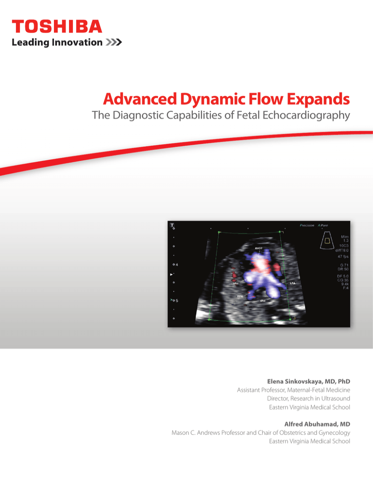
Toshiba’s Advanced Dynamic Flow (ADF), available on the Aplio™
Platinum series ultrasound systems, offers high-resolution,
color flow that enables physicians to identify particularly small
blood vessels and complex blood flow. ADF offers superior
spatial resolution at high frame rates to accurately depict flow
with directional information even in tiny vessels. Conducting a
fetal echocardiography examination using ADF enables a key
clinical evaluation during the early stages of a pregnancy. This
examination permits physicians to perform a comprehensive
evaluation of the baby’s cardiovascular system in utero, achieving
high-resolution imaging of the fetus’ heart, arteries, veins as well
as a study of blood flow. A thorough fetal echocardiographic
exam can identify potential anomalies during the early stages of a
pregnancy and help direct appropriate pregnancy management.
For the heart, we can obtain high-resolution snapshots of the
key anatomical structures using ADF (Figures 1-11). For example,
we can appreciate the four chambers and assess inlet blood
Figure 1: Transverse section of the fetal chest at the level of the four-chamber view obtained from a fetus at 16 weeks gestation showing normal
inlet blood flow.
dAo – descending aorta, RV – right ventricle, LV – left ventricle, RA – right atrium, LA – left atrium
Figure 2: Transverse section of the fetal chest at the level of the four-chamber view obtained from a fetus at 13 weeks gestation showing normal
inlet blood flow and intact interventricular septum.
RV – right ventricle, RA – right atrium, LV – left ventricle, LA – left atrium, dAo – descending aorta
2
Advanced Dynamic Flow Expands the Diagnostic Capabilities of Fetal Cardiography
flow, outflow tracts, and the interventricular septum. Blood flow
within the great vessels (aorta, pulmonary artery, pulmonary vein,
superior vena cava, inferior vena cava) is depicted in great detail.
In addition, the appearance of the atrial appendages, which
normally are not visible via traditional echocardiography is clearly
visualized using ADF. ADF permits a view into the development
of the heart, at a particularly early stage including during the first
trimester, which offers clinical benefits for the early detection of
fetal anomalies.
With the Aplio 500 Platinum, we can also visualize the
comprehensive renal system anatomy and vascular function in a
fetus, including the blood flow and branching details within the
main renal artery and vein and other intra-renal vessels (Figure 12).
Also, ADF can be used to image the fetal umbilicoportohepatic
system with great spatial resolution and accuracy (Figure 13).
ADF is an important tool that enables physicians to perform a
sophisticated fetal cardiovascular assessment of the fetal heart
Figure 3: ADF highlights the appearance of the right atrial appendages. (A) Right parasagittal section of the fetal chest obtained from a fetus at
13 weeks gestation showing the drainage of the superior and inferior caval veins into the right atrium. (B) Transverse section of the fetal chest
obtained from a fetus at 13 weeks gestation showing the four-chamber view.
RA – right atrium, SVC – superior vena cava, IVC – inferior vena cava, RV – right ventricle, LV – left ventricle, LA – left atrium
Figure 4: Transverse section of the fetal chest at the level of the four-chamber view obtained from a fetus at 16 weeks gestation showing the
drainage of the pulmonary veins into left atrium. ADF highlights the appearance of atrial appendages.
3
Advanced Dynamic Flow Expands the Diagnostic Capabilities of Fetal Cardiography
at the earliest possible stage of pregnancy, as early as 12 weeks.
physicians and patients the ability to detect many potential
Typically, such insights were not previously visible until week
health issues as early as possible. The clarity of the images
20 of pregnancy. Patients benefit from receiving these early
provides a distinct improvement in visualization of the fetal
insights, potentially giving them assurances that their gestation
anatomy. This ensures that physicians can evaluate more fully
is progressing normally. The benefits of ADF from the Aplio
the development process of a fetus and provide better care and
500 Platinum ultrasound system are wide-ranging, providing
more accurate treatment options for patients.
Figure 5: Sagittal section of the fetal chest obtained from a fetus at 16 weeks gestation showing the relation of the aortic and ductal arches (DA).
dAo – descending aorta, UV – umbilical vein, DV- ductus venosus, Ao isthmus – aortic isthmus, Int. ThA – internal thoracic artery
Figure 6: Transverse/oblique section of the fetal chest obtained from a fetus at 16 weeks gestation showing the relation of the
great vessels and outflow tracts.
4
Advanced Dynamic Flow Expands the Diagnostic Capabilities of Fetal Cardiography
Figure 7: Transverse section of the fetal
chest at the level of the three vessel
view obtained from a fetus at 16 weeks
gestation showing the right ventricular
outflow tract (RVOT) and bifurcation of
the pulmonary artery.
dAo – descending aorta, aAo – ascending
aorta, RPA- right pulmonary artery, LPA –
left pulmonary artery
Figure 8: Right parasagittal section of the fetal chest obtained from a fetus at 16 weeks gestation showing the drainage of the superior vena cava
(SVC) and inferior vena cava (IVC) into the right atrium (RA).
AzV – azygos vein
Figure 9: Transverse sections of the fetal chest at the level of the three vessel view (A) and three vessel trachea view (B) obtained from a fetus at 13
weeks gestation showing the relation of the great vessels.
PA – pulmonary artery, aAo – ascending aorta, RPA right pulmonary artery, LPA – left pulmonary artery, Tr- trachea, dAo – descending aorta, AzV – azygos vein
5
Advanced Dynamic Flow Expands the Diagnostic Capabilities of Fetal Cardiography
Figure 10: Transverse section of the fetal chest at the level of the three vessel trachea view obtained from a fetus at 16 weeks gestation showing
the aortic and ductal arches.
PA – pulmonary artery, Ao – aorta, SVC – superior vena cava, Tr – trachea, DA – ductal arch
Figure 11: Transverse oblique view of the upper fetal chest at the level of drainage of the left brachiocephalic vein (LBCV) into superior vena cava
(SVC) obtained from fetus at 26 weeks gestation.
Tr – trachea
6
Advanced Dynamic Flow Expands the Diagnostic Capabilities of Fetal Cardiography
Figure 12: Assessment of the main renal artery, main renal vein, and intra-renal vessels on a fetus at 32 weeks gestation using ADF.
Figure 13: Transverse section of the fetal upper abdomen at the level of the portal sinus obtained from a fetus at 32 weeks gestation. Normal fetal
umbilicoportohepatic system is imaged using ADF.
Ao – descending aorta, DV – ductus venosus, IVC – inferior vena cava, LPV – left portal vein, RPV – right portal vein, UV – umbilical vein
The clinical results described in this paper are the experience of the authors. Results may vary due to clinical setting, patient presentation and other factors.
ADF provides detailed visualization of blood flow with high frame rates.
7
Advanced Dynamic Flow Expands the Diagnostic Capabilities of Fetal Cardiography
Toshiba gives you a voice. What’s yours?
2441 Michelle Drive, Tustin CA 92780 / 800.421.1968 medical.toshiba.com
©Toshiba Medical Systems Corporation 2015. All rights reserved. Design and specifications are subject to change without notice.
Aplio is a trademark of Toshiba Medical Systems Corporation.
ULWP12442US
MCAUS0260EB


