Functional Anatomy Anatomical Position Descriptions of Directions 1/19/2016
advertisement
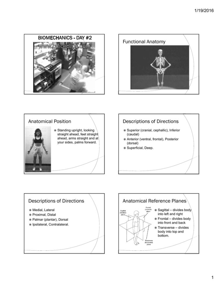
1/19/2016 Functional Anatomy Anatomical Position Standing upright, looking straight ahead, feet straight ahead, arms straight and at your sides, palms forward. Descriptions of Directions Medial, Lateral Proximal, Distal Palmar (plantar), Dorsal Ipsilateral, Contralateral. Descriptions of Directions Superior (cranial, cephallic), Inferior (caudal) Anterior (ventral, frontal), Posterior (dorsal) Superficial, Deep. Anatomical Reference Planes Sagittal – divides body into left and right Frontal – divides body into front and back Transverse – divides body into top and bottom. 1 1/19/2016 Descriptions of Motion Flexion, Extension Abduction, Adduction Elevation, Depression Internal rotation, External rotation Pronation, Supination Joint Planes of Action • Plantarflexion, Dorsiflexion • Lateral flexion • Radial deviation, ulnar deviation • Inversion, eversion • Circumduction. Types of Tissues Four tissue classifications Epithelial Nervous Muscle Connective. Epithelial Tissue Basically a covering, or lining, tissue Can be specialized to absorb, secrete, transport, excrete, or protect the underlying organ or tissue Nourished via tissue fluid from capillaries from connective tissues Plays a role in diffusion of fluid and heat. Epithelial Tissue Subject to wear - cells are constantly being lost and regenerated May exist as one or more layers Can be arranged in squamous, cuboidal, or columnar structures Not strong, typically bound to connective tissues. 2 1/19/2016 Nervous Tissue Comprises the main parts of the nervous system brain spinal cord peripheral nerves nerve endings Characteristics of Nervous Tissue Irritability - capacity to react to chemical or physical agents Conductivity - ability to transmit impulses from one location to another. sense organs. Nerve Cells Nerve Cells The neuron, or nerve cell, is the basic unit of nervous tissue. Cell body Axon The neuron, or nerve cell, is the basic unit of nervous tissue. Cell body Dendrites. Axon Dendrites Figure from “Structure and Function of the Musculoskeletal System”, Human Kinetics Nerve Cells Figure from “Structure and Function of the Musculoskeletal System”, Human Kinetics Muscle Tissue Nerve impulses are conducted toward the cell body by the dendrites and away from the cell body by the axon. Three Categories of Muscle Smooth Cardiac Skeletal. Figure from “Structure and Function of the Musculoskeletal System”, Human Kinetics 3 1/19/2016 Composition Aggregate of: cells fibres other macromolecules matrix tissue fluid. Fibres Fibre Arrangement and Density Types of Fibres loose connective tissue dense connective tissue ○ dense irregular connective tissue Collagen Reticular fascia Elastic. Loose Connective Tissues Four Types fibroelastic - encapsulates most organs areolar - saturates most every area of the body reticular - found near lymph nodes, and in bone marrow, liver, and spleen adipose - aggregate of fat cells. Arrangement and density of fibres distinguishes ○ dense regular connective tissue tendon, ligament, bone, etc. Constituents of Connective Tissues Resident Cells Undifferentiated mesenchymal cells ○ can differentiate into many different types of connective tissue cells ○ form fibres and other components of matrix. 4 1/19/2016 Constituents of Connective Tissues Extracellular Matrix Migratory cells travel to tissue via the bloodstream associated with tissues reaction to injury simple and complex glycoproteins part of major defense system of body Mast Cells - can transport heparin (anticoagulant), histamine (vasodilator), and serotonin (vasoconstrictor) to injured tissues. Proteoglycan Aggregate Blend of protein fibres (collagen and elastin) Macrophage - can assimilate foreign material, tissue fluid. Collagen Fibres Present in varying amounts in all connective tissues Can be arranged randomly or in a highly oriented fashion More than 20 different types of collagen Type I - skin, bone, tendon, ligament, cornea Type II - cartilage Type III - loose connective tissues. Collagen Fibres Hierarchy More slender and extensible than collagen Can be stretched to 150% of their original length Composed of microfibrils and elastin. tropocollagen molecule, aligned in rows to form microfibrils, arranged in bundles to form collagen fibres Elastic Fibres Molecules may be cross-linked which increases stiffness. 5 1/19/2016 Glycoproteins Tissue Fluid Occupy spaces between fibres Constitute the so-called “ground substance” of connective tissues Negatively charged and hydrophilic. Bone Components of Bone A filtrate of the blood Resides in the intercellular (interstitial) spaces Aids in transport between cells and capillaries. Osteoblasts - responsible for bone formation on surface of bone Osteocytes - formed when osteoblast surrounds itself with mineralized matrix, responsible for bone formation Osteoclasts - resorb bone Extracellular bone matrix. One of the hardest and strongest tissues of the body Protects organs, stores minerals, houses bone marrow, provides levers for muscles Dynamic structure, constantly remodeling itself in response to alterations in loading. Bone Cortical (also called compact) forms the hard, outer shell of bones Trabecular (also called cancellous or spongy). 6 1/19/2016 Development of Bone Development of Bone Can be influenced by movement and related forces during skeletal development Normal growth can be interrupted by trauma or fracture. Bone with damaged growth plate Fig 2.7 Bone Macrostructure 7 1/19/2016 Bone Microstructure Cartilage Hyaline - covers the surface of most joints Elastic - found in external ear, larynx, epiglottis, eustachian tube Fibrocartilage - found at stress points where friction could be a problem. Cartilage Hyaline Cartilage Has no intrinsic blood vessels, nerves, or lymph vessels Nutrients and wastes transported by diffusion. Articular Cartilage Tendons and Ligaments Made up of organized bundles of collagen fibres parallel in tendon parallel, oblique, or spiral in ligament Have great tensile strength Resist stretching in one direction. 8 1/19/2016 Tendons and Ligaments Tendon Hierarchy Can contain sensory receptors Ruffini corpuscles Pacinian corpuscles Golgi tendon organs free-nerve endings. Skeletal Muscle Muscle Structure Contain contractile proteins and connective tissue Serve as prime executors (movers) of the nervous system. Muscle Structure Muscle Structure 9 1/19/2016 Sliding Filament Theory Cross-Bridge Cycle Characteristics of Muscle Fibres Fibre Orientation Pennation effects the force that a muscle can develop as well as the amount that it can shorten. 10 1/19/2016 Motor Unit Length vs. Tension A motor unit is the basic of the neuromuscular system. Case Study: Knee and Ankle Sprains Force vs. Velocity Sprains are injuries to ligaments, usually due to sudden overstretching Typically associates with pain, inflammation, and loss of function Other impacts are not fully appreciated. Case Study: Knee and Ankle Sprains Many (all?) ligaments have nerve fibers in them These sensory receptors provide feedback that can trigger reflex muscle contraction to dynamically stabilize joints Loss of this feedback can lead to further instability and alterations in movement patterns. Case Study: Knee and Ankle Sprains Ankle Orthotics and Sprains Inversion sprains are common Previous sprains make subsequent ones more likely Commonly use taping or bracing to enhance stability and attempt to prevent reinjury Taping is costly, time and skill intensive, and has decreasing utility 11 1/19/2016 Case Study: Knee and Ankle Sprains Ankle Bracing Case Study: Knee and Ankle Sprains ACL Sprains Bracing is not fully evaluated Receptors in ACL have been Study examined soccer players, shown to provide feedback to the hamstrings Both the ACL and hamstrings serve to prevent anterior translation of the tibia relative to the femur Loss of ACL leads to instability, but also alterations in gait patterns. some of whom had previously suffered an ankle sprain and some who hadn’t Bracing was beneficial to those with previous injury but not those without Why? Arthrology Classification of Joints Classifications With a joint cavity ○ Diarthrodial Freely moveable Without a joint cavity ○ Synarthrodial Immovable ○ Amphiarthodial Slightly moveable. 12 1/19/2016 Knee Cross-Section 13
