AP Biology Animal Histology Lab
advertisement
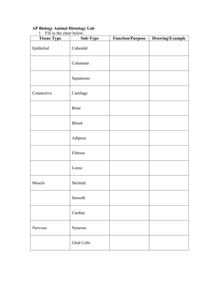
AP Biology Animal Histology Lab 1. Fill in the chart below: Tissue Type Sub-Type Epithelial Cuboidal Columnar Squamous Connective Cartilage Bone Blood Adipose Fibrous Loose Muscle Skeletal Smooth Cardiac Nervous Neurons Glial Cells Function/Purpose Drawing/Example 2. Examine the Bone slide. Answer the following: a. Draw what you see and label any cells that you can identify. b. What are the purposes of osteoblasts and osteons? c. What makes bone strong? 3. Examine the Blood slide. a. What cells can you draw and identify? b. Distinguish between the different components of blood (ex: plasma) 4. Examine the Nerve slide. a. Draw a nerve cell from the scope and label the axon, dendrites, synapse, cell body. b. In which direction does the nerve impulse travel? c. What’s the purpose of glial cells? 5. Examine the Columnar Epithelium slide. a. Where would you expect to find cells like this? b. What does it mean if the columnar cells are stratified? c. What is Transitional epithelium for? 6. Examine the Trachea slide. a. What kind of tissue is this made of? b. Where else would you expect to find this tissue? 7. Examine the Muscle slide. a. Draw and label each of the 3 types of muscle cells on this slide. b. Where would you expect to find each of these cells? c. Why are skeletal muscle cells multinucleate? 8. General Questions: a. What is the role of collagen, fibroblasts, and macrophages in connective tissue? b. Distinguish between collagenous, elastic, and reticular fibers.
