Current Research Journal of Biological Sciences 6(3): 117-121, 2014
advertisement
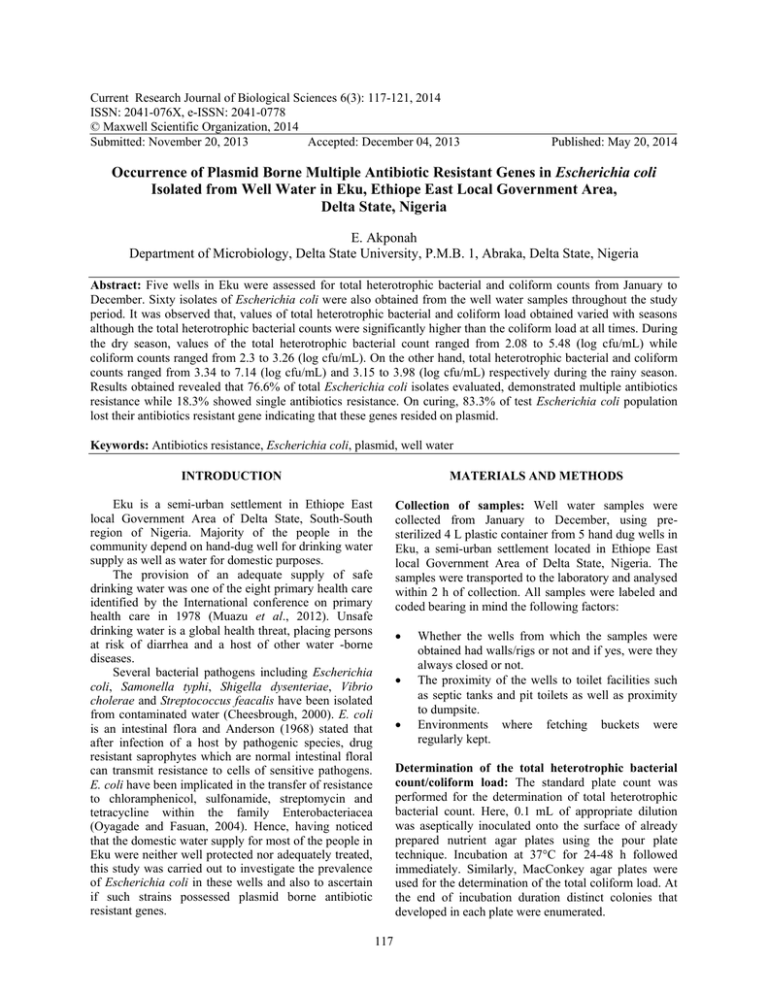
Current Research Journal of Biological Sciences 6(3): 117-121, 2014 ISSN: 2041-076X, e-ISSN: 2041-0778 © Maxwell Scientific Organization, 2014 Submitted: November 20, 2013 Accepted: December 04, 2013 Published: May 20, 2014 Occurrence of Plasmid Borne Multiple Antibiotic Resistant Genes in Escherichia coli Isolated from Well Water in Eku, Ethiope East Local Government Area, Delta State, Nigeria E. Akponah Department of Microbiology, Delta State University, P.M.B. 1, Abraka, Delta State, Nigeria Abstract: Five wells in Eku were assessed for total heterotrophic bacterial and coliform counts from January to December. Sixty isolates of Escherichia coli were also obtained from the well water samples throughout the study period. It was observed that, values of total heterotrophic bacterial and coliform load obtained varied with seasons although the total heterotrophic bacterial counts were significantly higher than the coliform load at all times. During the dry season, values of the total heterotrophic bacterial count ranged from 2.08 to 5.48 (log cfu/mL) while coliform counts ranged from 2.3 to 3.26 (log cfu/mL). On the other hand, total heterotrophic bacterial and coliform counts ranged from 3.34 to 7.14 (log cfu/mL) and 3.15 to 3.98 (log cfu/mL) respectively during the rainy season. Results obtained revealed that 76.6% of total Escherichia coli isolates evaluated, demonstrated multiple antibiotics resistance while 18.3% showed single antibiotics resistance. On curing, 83.3% of test Escherichia coli population lost their antibiotics resistant gene indicating that these genes resided on plasmid. Keywords: Antibiotics resistance, Escherichia coli, plasmid, well water INTRODUCTION MATERIALS AND METHODS Eku is a semi-urban settlement in Ethiope East local Government Area of Delta State, South-South region of Nigeria. Majority of the people in the community depend on hand-dug well for drinking water supply as well as water for domestic purposes. The provision of an adequate supply of safe drinking water was one of the eight primary health care identified by the International conference on primary health care in 1978 (Muazu et al., 2012). Unsafe drinking water is a global health threat, placing persons at risk of diarrhea and a host of other water -borne diseases. Several bacterial pathogens including Escherichia coli, Samonella typhi, Shigella dysenteriae, Vibrio cholerae and Streptococcus feacalis have been isolated from contaminated water (Cheesbrough, 2000). E. coli is an intestinal flora and Anderson (1968) stated that after infection of a host by pathogenic species, drug resistant saprophytes which are normal intestinal floral can transmit resistance to cells of sensitive pathogens. E. coli have been implicated in the transfer of resistance to chloramphenicol, sulfonamide, streptomycin and tetracycline within the family Enterobacteriacea (Oyagade and Fasuan, 2004). Hence, having noticed that the domestic water supply for most of the people in Eku were neither well protected nor adequately treated, this study was carried out to investigate the prevalence of Escherichia coli in these wells and also to ascertain if such strains possessed plasmid borne antibiotic resistant genes. Collection of samples: Well water samples were collected from January to December, using presterilized 4 L plastic container from 5 hand dug wells in Eku, a semi-urban settlement located in Ethiope East local Government Area of Delta State, Nigeria. The samples were transported to the laboratory and analysed within 2 h of collection. All samples were labeled and coded bearing in mind the following factors: • • • Whether the wells from which the samples were obtained had walls/rigs or not and if yes, were they always closed or not. The proximity of the wells to toilet facilities such as septic tanks and pit toilets as well as proximity to dumpsite. Environments where fetching buckets were regularly kept. Determination of the total heterotrophic bacterial count/coliform load: The standard plate count was performed for the determination of total heterotrophic bacterial count. Here, 0.1 mL of appropriate dilution was aseptically inoculated onto the surface of already prepared nutrient agar plates using the pour plate technique. Incubation at 37°C for 24-48 h followed immediately. Similarly, MacConkey agar plates were used for the determination of the total coliform load. At the end of incubation duration distinct colonies that developed in each plate were enumerated. 117 Curr. Res. J. Biol. Sci., 6(3): 117-121, 2014 Isolation and identification of Escherichia coli: Lactose fermenting colonies on the MacConkey plates were randomly sub-cultured into Eosin methylene blue agar plates and incubation followed at 37°C for 18-24 h. Colonies with green metallic sheen colour suggestive of E. coli were furthermore subjected to biochemical characterization according to criteria in Cruickshank et al. (1975) for confirmation. Sodium Dedoecyl Sulphate (SDS) solution in a ratio of 4:1. Furthermore, the mixture was incubated at 42°C for 2 h. After curing, the cells were harvested, washed with sterile deionized water and standard inocula obtained were tested for antibiotic sensitivity as described in the section above. Evaluation of the antibiotic susceptibility pattern of E. coli isolates to various antibiotics: The KirbyBauer NCCLS modified disc diffusion technique described in Cheesebrough (2000), was adapted for both antibiotic sensitivity assay and interpretations of zones of inhibition. The bacterial inoculum was standardized by inoculating 5 mL of freshly prepared (sterile) Nutrient broth with 3 colonies of 18-24 h culture of E. coli. The broth was then incubated at 37°C until its turbidity matched that of 1.0% BaSO 4 . The standardized inoculum was inoculated onto freshly prepared nutrient agar plates by swabbing the surface of the plate with swab stick soaked with the standard inoculum. The plates were allowed to stand for 3-5 min in order to absorb excess fluid. Subsequently, the multiple antibiotics disc was aseptically placed on the seeded agar plates and incubation followed at 37°C for 18-24 h. At the end of incubation period, zones of inhibition were measured and recorded. The antibiotics disc used were obtained from Maxicare laboratory, Lagos, Nigeria. It comprised of the following: septrin (30 mg), ampicillin (30 mg), ceporex (10 mg), ciprofloxacin (10 mg), nalidixic acid (30 mg), augumentin (30 mg), gentamicin (10 mg), perflaxin (10 mg), tarvid (10 mg) and streptomycin (10 mg). The total heterotrophic bacterial and coliform counts obtained varied with season. Elevated counts for both total heterotrophic bacterial count as well as total coliform counts were recorded in months ranging from June to September. Results of the total heterotrophic bacterial counts and total coliform load of well water samples obtained from the five stations are presented in Fig. 1 and 2 respectively. Generally, the coliform counts obtained in each month were lower than the total heterotrophic bacterial count. There was a significant difference at p≤0.05 between values of total heterotrophic bacterial counts and total coliform count from January to December Sixty E. coli isolates were obtained and 80% of which demonstrated resistant to nalidixic acid. Also, of the 60 E. coli strains, 75, 50, 40, 26.67, 25, 0, 3.33, 45 and 0(%) demonstrated resistance to septrin, streptomycin, gentamycin, ceporex, ampicillin, perflacin, augumentin, ciprofloxacin and tarvid respectively as shown in Table 1. However, 15% (3 isolates) were susceptible to all antibiotics used, 18.3% (11 isolates) showed single antibiotics resistance while 76.67% showed multiple antibiotics resistance as also indicated in Table 1. At the end of curing exercise which involved 57 E. coli strains of the total isolates obtained, it was observed that majority of the antibiotic resistant genes present resided in plasmids as 83.3% of tested population (50 isolates) lost resistance to various antibiotics. Results of the antibiogram of cured isolates are illustrated in Table 2. Statistical analysis (using the Student t-test) showed that there was a significant difference in the zones of inhibition produced by each antibiotics to each isolate before and after curing of the isolate (p≤ 0.05). RESULTS Log (cfuml-1) of total heterotrophic bacterial count Curing of isolates: Isolates that showed resistance to any of the antibiotics listed, were subjected to the curing experiment to determine if the gene responsible for such antibiotics resistant was resident on plasmid or not. A loopful of the isolate was aseptically introduced into sterile freshly prepared nutrient broth and incubation at 37°C followed for 24 h. At the end of incubation, the broth culture was introduced into 10% 8 7 6 5 4 3 2 1 0 WELL 1 WELL2 WELL3 Month of sampling Fig. 1: Seasonal changes in total heterotrophic bacterial count in various well water 118 WELL4 WELL5 log (cfuml-1)of total coliform count Curr. Res. J. Biol. Sci., 6(3): 117-121, 2014 WELL 1 WELL2 WELL3 WELL4 WELL5 4.5 4 3.5 3 2.5 2 1.5 1 0.5 0 Month of sampling Fig. 2: Seasonal changes in total coliform count in various well water Table 1: Antibiotic resistance pattern of Escherichia coli isolates to various antibiotics prior to curing Nalidixic acid Septrin Streptomycin Gentamycin Ceporex Ampicillin Isolate ISW1 a S S S S R S ISW1 b S R R R S R ISW1 c R R R R S S ISW1 d R R R R S S ISW1 e S S S S S S ISW1 F R R S R R R ISW1 G R R R R S R ISW1 H R R R R R R ISW1 I S R S R R S ISW1 J R R S R R S ISW1 K R R R S S S ISW1 L R R R S S S ISW2 A R R S S S S ISW2 B R R S R S S ISW2 C R R S S S S ISW2 D R R S R S R ISW2 E R R R R S S ISW2 F S S R S S S ISW2 G R S S S S S ISW2 H R R R R S R ISW2 I S R R R S R ISW2 J R R R S R R ISW2 K R R R R R S ISW2 L R S S S S S ISW3 A S S S S S S ISW3 B S S S S S S ISW3 C S S S S S S ISW3 D R R R S S S ISW3 E R R R S S S ISW3 F R S S S S S ISW3 G R S S S S S ISW3 H R S S S S S ISW3 I R R S R R R ISW3 J R R S S R S ISW3 K R R S S R S ISW3 L S R S S R S ISW4 A R R R S S R ISW4 B R R R S S S ISW4 C R S S S S S ISW4 D R R R R S S ISW4 E S R R R R S ISW4 F R R R R S R ISW4 G R R S R S S ISW4 H R R S R R R ISW4 I R R S S S S ISW4 J R R S S S R ISW4 K R R R S R S ISW4 L R R R S S S ISW5 A R S S S S S ISW5 B R R R S S S ISW5 C R R R S S S ISW5 D R R S S S S ISW5 E R R S S S S ISW5 F R R S R S R ISW5 G R R R S R S ISW5 H R R R R S R ISW5 I R S S S S S ISW5 J R R R S R S ISW5 K S R R R S S ISW5 L R S R R S S Key: S = Sensitive, R = Resistant 119 Percflacin S S S S S S S S S S S S S S S S S S S S S S S S S S S S S S S S S S S S S S S S S S S S S S S S S S S S S S S S S S S S Augumentin S S S S S S R S S S S S S S S S S S S S S S S S R S S S S S S S S S S S S S S S S S S S S S S S S S S S S S S S S S S S Ciprofloxacin S R S S S S R S R R S R R S R S R S S S R R S S S S S S R S S S R S R R S S S R S R R R R R S S S S R S R R R R S R R S Tarvid S S S S S S S S S S S S S S S S S S S S S S S S S S S S S S S S S S S S S S S S S S S S S S S S S S S S S S S S S S S S Curr. Res. J. Biol. Sci., 6(3): 117-121, 2014 Table 2: Antibiotic resistance pattern of Escherichia coli isolates to various antibiotics after curing Nalidixic Isolate acid Septrin Streptomycin Gentamycin Ceporex Ampicillin ISW1 a S S S S R S ISW1 b S S S S S S ISW1 c S S S S S S ISW1 d R R R R S S ISW1 e S S S S S S ISW1 F S S S R R R ISW1 G S S S S S S ISW1 H S S S S S S ISW1 I S S S S S S ISW1 J S S S S S S ISW1 K S S S S S S ISW1 L S S S S S S ISW2 A S S S S S S ISW2 B S S S S S S ISW2 C S S S S S S ISW2 D S S S S S S ISW2 E S S S S S S ISW2 F S S S S S S ISW2 G S S S S S S ISW2 H R R R R S R ISW2 I S S S S S S ISW2 J S S S S S S ISW2 K S S S S S S ISW2 L S S S S S S ISW3 A S S S S S S ISW3 B S S S S S S ISW3 C S S S S S S ISW3 D S S S S S S ISW3 E S S S S S S ISW3 F S S S S S S ISW3 G R S S S S S ISW3 H R S S S S S ISW3 I S S S S S S ISW3 J S S S S S S ISW3 K S S S S S S ISW3 L S S S S S S ISW4 A S S S S S S ISW4 B S S S S S S ISW4 C S S S S S S ISW4 D S S S S S S ISW4 E S S S S S S ISW4 F S S S S S S ISW4 G S S S S S S ISW4 H S S S S S S ISW4 I S S S S S S ISW4 J R R S S S R ISW4 K S S S S S S ISW4 L S S S S S S ISW5 A S S S S S S ISW5 B S S S S S S ISW5 C S S S S S S ISW5 D S S S S S S ISW5 E S S S S S S ISW5 F S S S S S S ISW5 G S S S S S S ISW5 H S S S S S S ISW5 I S S S S S S ISW5 J S S S S S S ISW5 K S S S S S S ISW5 L S S S S S S S = Sensitive, R = Resistant Percflacin S S S S S S S S S S S S S S S S S S S S S S S S S S S S S S S S S S S S S S S S S S S S S S S S S S S S S S S S S S S S Augumentin S S S S S S S S S S S S S S S S S S S S S S S S S S S S S S S S S S S S S S S S S S S S S S S S S S S S S S S S S S S S Ciprofloxacin S S S S S S S S S S S S S S S S S S S S S S S S S S S S S S S S S S S S S S S S S S S S S R S S S S S S S S S S S S S S Tarvid S S S S S S S S S S S S S S S S S S S S S S S S S S S S S S S S S S S S S S S S S S S S S S S S S S S S S S S S S S S S bodies during the rainy season (Oyagade and Fasuan, 2004). The consistent occurrence of E. coli in various water samples is an indication of fecal pollution. All of the wells studied were not properly protected. Well 1 had no wall, Well 2 had just a rig of wall on top and the other 2 were completely walled but none had cover. These were representatives of the wells commonly found in the community which also might have subjected them to run offs from either surrounding dumpsite, accidental or deliberate dropping of materials including fetching buckets as well as wash-off containing fecal matter especially those of children and DISCUSSION Data obtained in this study showed variation in total heterotrophic bacterial and coliform counts throughout the year. The variations observed may be due to variation in seasons as months with high rainfall recorded higher total heterotrophic bacterial count as well as total coliform counts. During the raining season or in months in which the amount of rainfall were high, a corresponding increase in counts were noticed, probably because there were increased run offs or perhaps the total organic matter and dissolved salts in various wells increased which is characteristics of water 120 Curr. Res. J. Biol. Sci., 6(3): 117-121, 2014 domestic animals passed or disposed indiscriminately. Also, most of the wells studied were dug less than 10 m away from toilet facilities especially septic tanks, as the common practice in the community is buildings clustered either to accommodate expanding population or to maximize available land space.) Results obtained indicated that 57 of the 60 E. coli strains showed resistance to all antibiotics employed with the exception of perflacin and tarvid. These antibiotics and others such as augumentin to which percentage of resistant strains were low, are second line antibiotics which are expensive and therefore, may not be affordable by the indigenes thereby limiting abuse. The cheapness and availability of first line antibiotics as tested herein, may have encouraged their use or misuse and hence, might be responsible for the resistance displayed by a high percentage of test isolates observed in this study. Several authors such as Bucknell et al. (1997), Watkinson et al. (2007) and Oyagade and Fasuan (2004), had shown that E. coli is a significant reservoir of genes coding for antimicrobial drug resistance. The result obtained in this study is at par with their results and thus is suggestive that E. coli can serve as useful indicator for acquired resistance in bacterial communities. The results of the curing experiment revealed that majority of E. coli isolates carried the antibiotics genes on transmissible plasmids. This could facilitate both intra and inter species transfer and in turn create a new level of risk associated with antibiotics resistance among the members of Eku community that depend on hand-dug wells for domestic water supply. It is therefore of paramount importance to organize campaigns to enlighten members of the community on possible or locally achievable water treatment procedures. Government is also, hereby, called upon to take provision of portable water supply more seriously especially in the Eku community of Delta State, Nigeria. REFERENCES Anderson, E.S., 1968. The ecology of transferable drug resistance in bacteria. Ann. Rev. Microbiol., 22: 131-180. Bucknell, D.G., R.B. Gasser, A. Frring and K. Whithear, 1997. Antimicrobial drug resistance in Salmonella and Escherichiacoli isolated from horses. Aust. Vet. J., 75: 355-356. Cheesebrough, M., 2000. District Laboratory Practice in Tropical Countries. Part 2. Cambridge University Press, UK, pp: 434. Cruickshank, R.J., P. Dugid, B.P. Marmuon and R.H.A. Swain, 1975. Medical Microbiology. 12th Edn., Churchill Livingstone, London, pp: 426-437. Muazu, J., A. Muhammad-Biu and G.T. Mohammed, 2012. Microbial quality of packaged sachet water marketed in Maiduguri metropolis, Nort Eastern Nigeria. Brit. J. Pharm. Toxicol., 3(1): 33-38. Oyagade, J.O. and O.O. Fasuan, 2004. Incidence of multiple antibiotic resistant Escherichia coli isolated from well water in Ado-Ekiti Nigeria. Nig. J. Microbiol., 18(1-2): 256-261. Watkinson, A.J., G.R. Micalizzi, J.R. Bates and S.D. Constanzo, 2007. Novel method for rapid assessment of antibiotic resistance in Escherichia coli isolates from environmental waters by use of a modified chromogenic agar. Appl. Environ. Microb., 73(7): 2224-2229. 121
